Organoid > Volume 2; 2022 > Article
Mun, Lee, Shin, Eun, Ji, and Son: Recent advances in multicellular human liver models

Abstract

The liver is the most important metabolic organ in the body. Model systems that recapitulate the complex organ structure and cell composition of the human liver are insufficient to study liver biology and to test toxicity and efficacy during new drug development. Recently established 3-dimensional liver models, including spheroids and organoids, organs-on-a-chip, bioprinting, and the decellularization/recellularization technique, have provided platforms that emulate the structural and functional characteristics of the human liver better than conventional 2-dimensional cell culture models and animal models. This review summarizes the architecture and cell compositions of human liver tissue, focusing on recent studies of multicellular human liver models that recapitulate in vivo-like physiologies with morphological and functional advances by the cellular communication of parenchymal and non-parenchymal cells. We discuss the applications, limitations, and future perspectives of advanced multicellular human liver models.

Introduction

The liver is a central organ in various metabolic processes, and its primary functions are (1) metabolism of carbohydrates, lipids, amino acids, and bile acids; (2) detoxification; (3) synthesis of plasma proteins such as albumin and clotting factors; and (4) storage of glycogen, vitamins, and minerals [1]. Hepatic failure arises due to the deterioration of liver function, mainly owing to xenobiotics or disease progression, and is often fatal. Unfortunately, reliable liver models for predicting the risk of hepatotoxicity during new drug development and evaluating the efficacies of drugs to target liver diseases are insufficient.
Three-dimensional (3D) liver models, including spheroids and organoids, have recently been developed, and their advantages were highlighted with respect to recapitulation of the complexities of cell compositions and tissue structure in comparison with 2-dimensional (2D) cell culture models. Primary human hepatocytes (PHHs) isolated from human liver tissue have high functionality and are therefore generally used as the gold standard for an in vitro drug testing platform. However, their viability and functionality are not sufficiently maintained in conventional 2D culture formats. Approaches to manage the 3D microenvironment by controlling stiffness using extracellular matrix (ECM), such as Matrigel and hydrogel, have been implemented and improved the viability of PHHs until 40 days in culture [2,3]. In addition, interactions between hepatocytes and non-parenchymal cells, such as endothelial cells and hepatic stellate cells (HSCs), in 3D hepatic spheroids improved viability and the liver phenotype [4,5]. Importantly, ameliorated liver functions, including high cytochrome P450 (CYP) enzyme activities, improved toxicity prediction in these 3D liver models.
As an alternative source of hepatic cells, protocols have been developed to differentiate induced pluripotent stem cells (iPSCs) into liver tissue cells, including hepatocytes [6], HSCs [7], and Kupffer cells (KCs) [8]. Furthermore, organoid generation protocols have been developed by using a 2D differentiation protocol in combination with 3D culture systems to provide a tissue microenvironment. Liver organoids have been generated from adult human liver tissue and remain stable and functional for a long time [9]. However, tissue-derived organoids mainly contain parenchymal cell types of liver tissue, such as hepatocytes and cholangiocytes, under epithelial cell type-enriched culture conditions. Therefore, iPSC-derived hepatic endoderm cells were co-cultured with endothelial cells and mesenchymal stem cells (MSCs) [10]. Furthermore, iPSC-based liver organoids containing multi-lineage cells were developed depending on their pluripotency [11]. This review summarizes the structures and cell types of human liver tissue and in vivo-mimicking liver models, specifically focusing on the recent advances and future applications of multicellular human liver models with potential use for investigating liver diseases and testing promising therapeutic drugs.
Ethics statement: This study was a literature review of previously published studies and was therefore exempt from institutional review board approval.

Liver architecture and cell composition

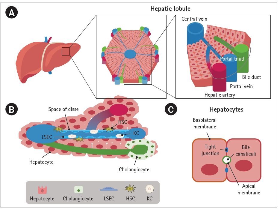
The basic architectural unit of the liver is the hepatic lobule, which consists of hepatocyte plates lined by sinusoidal capillaries radiating toward the central vein (Fig. 1A). Hepatic lobules are nearly hexagonal and each is delimited by the portal triad, consisting of the portal vein, hepatic artery, and bile duct. The portal vein and hepatic artery provide blood to the lobules through a sinusoidal capillary network from the portal triad to the central vein, while bile synthesized by hepatocytes flows in the opposite direction to the bile duct. Hepatocytes are the main parenchymal cell type of the liver and account for approximately 70% of liver cells and most of the liver volume [12]. Cholangiocytes and non-parenchymal stromal cells, including liver sinusoidal endothelial cells (LSECs), HSCs, and KCs are also important components of the liver (Fig. 1B and Fig. 2) [13-28].
Hepatocytes are positioned with their basolateral surface facing perforated LSECs, which facilitates the delivery of endocrine secretions into the bloodstream (Fig. 1C). Bile acids and bile salts are transported across the bile canalicular-apical surface of hepatocytes, and the basolateral and apical membranes are separated by tight junctions between adjacent hepatocytes [29]. The arrangement between polarized hepatocytes and capillaries and bile duct cells is associated with the endocrine and exocrine functions of the liver [30].
Cholangiocytes are the other parenchymal cells in the liver. They are highly specialized epithelial cells that line the biliary tree, a network of bile ducts, and account for approximately 3% to 5% of liver cells [22,28]. The major physiological functions of cholangiocytes are bile secretion, modification, and homeostasis through transport of various ions and water in a complex liver anatomic niche [31]. They also play a central role in liver regeneration in pathologic conditions [32].
LSECs are specialized hepatic endothelial cells that line the hepatic sinusoids, a capillary network in the liver, and are the most abundant non-parenchymal cells in the liver, accounting for approximately 15% to 20% of liver cells [33,34]. They are one of the most permeable endothelial barriers, allowing an easy exchange of metabolites and maintenance of homeostasis within the liver [16]. LSECs are also a selective barrier positioned at the interface between the oxygen- and nutrient-rich blood side and abluminal side with hepatocytes and HSCs; they are crucial for nutrient exchange, active uptake, the metabolism of proteins, lipids, and glucose, and the clearance of waste biomolecules and xenobiotics including drugs [34].
KCs are tissue-resident macrophages in the liver sinusoids and are the first line of immune cells that respond to infectious materials transported from the gastrointestinal tract via the portal vein [35]. Their major functions are involved in host systemic defense under physiologic conditions and in disease progression and resolution under pathologic conditions through phagocytosis, immunomodulation, and metabolic functions [36]. Liver injuries that overwhelm the gatekeeping function of KCs result in inflammation, fibrosis, and eventually hepatocellular carcinoma; therefore, KCs are important target cells for the treatment of liver diseases [37].
HSCs are positioned in the space of Disse, the perisinusoidal space between a hepatocyte and sinusoid (Fig. 1B). They mainly store vitamin A in a quiescent state and play important roles in regulating blood flow and maintaining the ECM balance by acting as pericyte-like cells in the liver [38]. Various insults that cause liver injury activate HSCs to transdifferentiate into myofibroblast-like cells, leading to collagen accumulation and fibrosis [39,40].

In vivo-mimicking liver models

1. 3D co-culture

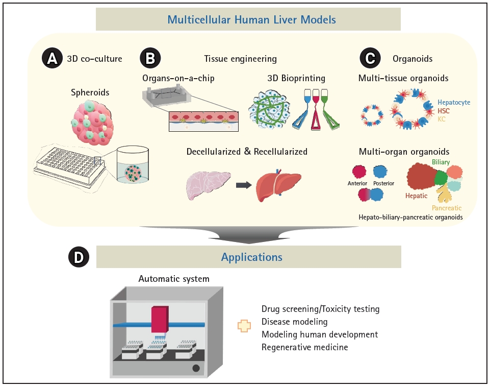
There is no perfect in vivo model of the liver; however, several in vitro multi-lineage liver models have been extensively developed (Fig. 3 and Table 1) [5,10,11,30-32,35-38,41-55]. Spheroids, which are 3D cultures of hepatic parenchymal and non-parenchymal cells, are among the most commonly used liver models [56] (Fig. 3A). Spheroids consisting of PHHs and non-parenchymal cells demonstrated improved metabolic function and stability in culture; thus, long-term drug treatment could be evaluated for drug-induced liver injury (DILI) [5]. Importantly, the sensitivity of 3D hepatic spheroids to known hepatotoxicants and their predictive value were better than those of plated 2D PHHs [5]. Moreover, spheroids are relatively homogeneous, easy to culture, and cost-effective; thus, they may be a more suitable model for high-throughput screening (HTS) than the more complex model systems described next. However, they show lower structural complexity and poorer in vivo fidelity than native tissue, which remain as limitations.

2. Tissue engineering

While spheroids primarily emulate 3D cell-cell interactions, tissue engineering technologies can provide the surrounding microenvironment to recapitulate the microarchitecture of native tissue and mechanical properties such as blood flow. Organs-on-a-chip system can simulate the positional specificity of each cell type and their spatial relationships (Fig. 3B). Polarized hepatocytes separated by a lining of endothelial cells to mimic the space of Disse generated using fluidics exhibited long-term survival and increased CYP enzyme activities [50,55]. Bioprinting technology can also provide micro-architectural properties using bioink-containing cells, growth factors, and biomaterials (Fig. 3B). Bioprinted liver models inspired by the liver lobular structure have been constructed. A liver ECM-based bioink has been designed to encapsulate liver cells and thereby mimic natural tissue properties, and cords of hepatocytes with a functional sinusoidal lumen-like network in both horizontal and vertical orientations were designed. The bioprinted liver model demonstrated enhanced albumin production, urea synthesis, and CYP enzyme activities [46].
An important element of 3D modeling is the types and concentrations of ECM molecules, which are essential for cell attachment, proliferation, and differentiation, and are mainly proteins, including collagens, elastin, and fibronectin, polysaccharides, and proteoglycans [57,58]. Decellularization of tissues or organs is a good method to prepare ECM that potentially maintains the natural microenvironment and architecture of the tissue [44,47] (Fig. 3B). Recellularized rat liver successfully preserved the survival and function of hepatocytes in vitro, and, upon transplantation of recellularized liver grafts into rats, the micro-anatomical organ structure, including the vascular network and biliary tract, was maintained and the function was similar to that of the adult liver [54]. Moreover, decellularized ECM can improve the hepatic maturation of stem cells. Bone marrow-derived MSCs cultured in a decellularized liver scaffold as spheroids exhibited mature and stable metabolic functions compared with those cultured in a 2D system. Notably, liver-specific ECM provides an improved environment for hepatic differentiation and maturation of stem cells [41].

3. Organoids

Organoids are stem cell-based self-organized 3D models that recapitulate the cell composition and physiological niche of the corresponding tissue [59]. Liver organoids can be generated from adult tissue stem cells [9,60] and pluripotent stem cells (PSCs) [53,61]. While tissue-derived organoids are generated in a relatively short amount of time and are genetically stable even after long-term passage [9], they have limited characteristics and differentiation potential owing to the characteristics of the cell source [62]. Meanwhile, PSCs, including embryonic stem cells and iPSCs, can differentiate into the three germ layers (ectoderm, mesoderm, and endoderm), which can develop into all types of cells and tissues. Therefore, PSCs can generate multi-lineage organoids from a single donor.
Multi-tissue organoids composed of cells of multiple germ layers within a single organ can be generated by intra-organ self-organization [63]. Multi-organ organoids can be generated by inter-organ self-organization through interconnectivity between organ domains [63] (Fig. 3C). Multi-tissue “liver bud” organoids have been generated by co-culturing cells derived from different germ layers, such as hepatic endoderm cells derived from iPSCs, human umbilical vein endothelial cells, and MSCs [10]. Liver buds composed of cells derived entirely from iPSCs were further developed [52]. Multi-tissue organoids can be used to examine multi-lineage communication and were used to reveal that vascular endothelial growth factor signaling crosstalk between hepatoblasts and endothelial cells strengthens endothelial network formation and hepatic differentiation [43]. When liver buds were transplanted into the kidney capsule or cranial window in vivo, they connected to the host vessel within 48 hours and matured to show functionality similar to that of the adult liver [43,53]. Furthermore, co-differentiation methods have been reported [11,45,51], and liver organoids consisting of hepatocyte-like cells, biliary cells, HSCs, and KCs were generated from iPSCs [11]. Single-cell RNA sequencing revealed that the model contained about 59.2% hepatocyte-like cells, 31% stellate-like cells, 8.9% biliary-like cells, and 0.8% Kupffer-like cells. Steatohepatitis has been successfully modeled with oleic acid (OA) treatment, and progressive disease phenotypes, such as increased lipid accumulation, triglyceride storage, inflammatory signaling, and stiffness, which represent the severity of fibrosis, were observed in OA-treated multicellular liver organoids [11]. In addition, high-fidelity DILI prediction was possible by evaluating cholestatic and/or mitochondrial toxicity using established organoids [51]. Another multi-lineage organoid model was developed for liver fibrosis, and these organoids have a ductal and vascular structure and contain mesenchymal cells [45]. Congenital hepatic fibrosis has been successfully modeled by engineering the causative mutation into organoids, and collagen-producing myofibroblasts, which are essential mediators of liver fibrosis, and the platelet-derived growth factor receptor beta (PDGFRB)-STAT3 pathway were activated. To this end, treatment with PDGFRB inhibitors elicited an anti-fibrotic effect in the organoid model of liver fibrosis [45]. These multi-tissue organoids faithfully emulate complex features of disease progression and can be used to evaluate toxicity and develop novel therapies. Finally, multi-organ organoids containing hepato-biliary-pancreatic organ domains have been established, followed by human endoderm organogenesis [48,64]. Hepato-biliary-pancreatic tissues developed at the boundary between anterior and posterior gut spheroids generated by iPSCs [48]. Retinoic acid signaling from non-epithelial cells in the anterior region at the foregut-midgut boundary was important for lineage specification [49]. Structurally and functionally integrated multi-organ organoids can be valuable models for performing mechanistic studies of complex organogenesis specifically in humans, modeling congenital disorders and related diseases, and developing therapeutic applications.

Applications, limitations, and future perspectives

Multicellular liver models including spheroids, organoids, and various 3D model systems in combination with tissue engineering technologies are useful alternatives to traditional in vitro and animal models. They have been used for diverse applications from basic research to therapeutic development, including modeling of human development and diseases, drug screening, toxicity testing, and regenerative medicine (Fig. 3D). Multicellular systems can model spatiotemporal cell-cell and cell-ECM communications because they recapitulate the structural and functional complexity of the liver tissue, which is difficult to achieve with 2D models. Notably, organoids can be generated using iPSCs or patient-derived cells, which may provide patient-specific models for the evaluation of differences in patients’ responses to personalized medicine. However, 3D modeling is time-consuming, labor-intensive, and less compatible with HTS, and these disadvantages are amplified according to the complexity of the physiological niche being mimicked [52,65]. An automatic platform has been adopted for robust and consistent generation, culture, and drug testing of organoids. Quality control and reproducibility are challenging owing to the heterogeneity of the models, and higher resolution techniques such as single-cell RNA sequencing and multi-omics are required to characterize multicellular systems. Chemically defined ECM and cost-efficient processes for large-scale manufacturing remain to be developed. The recent advances in multicellular human liver models may provide promising systems for biomedical applications and, ultimately, organ replacement.

NOTES

Conflict of interest

No potential conflict of interest relevant to this article was reported.

Funding

This work was supported by the Korea Research Institute of Bioscience and Biotechnology (KRIBB) Research Initiative Program (KGM4722223 and KGM5362212), a National Research Foundation (NRF) grant funded by the Korean government (MSIT) (NRF-2022R1A2B5B02001644), and a grant (22213MFDS386) from the Ministry of Food and Drug Safety, Korea, in 2022.

Data availability

Please contact the corresponding author for data availability.

Fig. 1.
Liver architecture and cell composition. (A) Structural organization of the liver, (B) Cell localization in the hepatic lobule, (C) Apicobasal membrane polarity in hepatocytes. LSEC, liver sinusoidal endothelial cell; HSC, hepatic stellate cell; KC, Kupffer cell.
organoid-2022-2-e26f1.jpg
Fig. 2.
Cell types of the liver and their major functions and representative markers. ALB, albumin; ACTA2: actin alpha 2, smooth muscle; HNF4A, hepatocyte nuclear factor 4 alpha; KRT, keratin; SOX9, SRY-Box transcription factor 9; LYVE1, lymphatic vessel endothelial hyaluronan receptor 1; STAB2, stabilin 2; ID I/III, inhibitor of DNA binding 1/3; CLEC-4F, C-type lectin domain family 4 member F; COL1A1, collagen type I alpha; PDGFRβ, platelet-derived growth factor receptor beta.
organoid-2022-2-e26f2.jpg
Fig. 3.
In vivo-mimicking multicellular human liver models and their applications. (A) tissue engineering, (C) organoids, (D) applications.
organoid-2022-2-e26f3.jpg
Table 1.
In vivo-mimicking multicellular liver models
Approaches Features Common advantages Common limitations Reference
3D co-culture
 PHHs/KCs Enhanced DILI sensitivity ∙ Easy to culture ∙ Structural complexity↓ [5]
∙ Suitable for HTS ∙ Low in vivo similarity
 PHHs/LSECs/KCs /HSCs Enhanced hepatic functions ∙ Relatively homogeneous [42]
Tissue engineering
 Organs-on-a-chip Spatial relationships with fluidics In vivo-mimicking of the microenvironment ∙ Less compatible with HTS [30,31]
 Bioprinting Enhanced structural complexity ∙ Tissue-like structures ∙ Comparatively difficult to culture [32]
 Decellularized scaffolds Native tissue structures, tissue-specific ECM [35-38]
Organoids
 Multi-tissue organoids Liver buds (vascularization/easy engrafting in vivo) ∙ Structural complexity↑ ∙ High cost [10,41,45,46]
∙ Long-term stability ∙ Time-consuming
∙ Cryopreservation ∙ Less compatible with HTS
Co-differentiation (advanced disease modeling and DILI prediction) ∙ High fidelity [11,47,48]
 Multi-organ organoids Integrated hepato-biliary-pancreatic tissues ∙ Patient-specific models [49,51]

3D, 3-dimensional; PHH, primary human hepatocyte; KC, Kupffer cell; DILI, drug-induced liver injury; HTS, high-throughput screening; LSEC, liver sinusoidal endothelial cell; HSC, hepatic stellate cell; ECM, extracellular matrix.

References

1. Trefts E, Gannon M, Wasserman DH. The liver. Curr Biol 2017;27:R1147-51.
crossref pmid pmc
2. Lee HJ, Son MJ, Ahn J, Oh SJ, Lee M, Kim A, et al. Elasticity-based development of functionally enhanced multicellular 3D liver encapsulated in hybrid hydrogel. Acta Biomater 2017;64:67-79.
crossref pmid
3. Lee HJ, Ahn J, Jung CR, Jeung YJ, Cho HS, Son MJ, et al. Optimization of 3D hydrogel microenvironment for enhanced hepatic functionality of primary human hepatocytes. Biotechnol Bioeng 2020;117:1864-76.
crossref pmid pdf
4. Foster AJ, Chouhan B, Regan SL, Rollison H, Amberntsson S, Andersson LC, et al. Integrated in vitro models for hepatic safety and metabolism: evaluation of a human Liver-Chip and liver spheroid. Arch Toxicol 2019;93:1021-37.
crossref pmid pdf
5. Proctor WR, Foster AJ, Vogt J, Summers C, Middleton B, Pilling MA, et al. Utility of spherical human liver microtissues for prediction of clinical drug-induced liver injury. Arch Toxicol 2017;91:2849-63.
crossref pmid pmc pdf
6. Si-Tayeb K, Noto FK, Nagaoka M, Li J, Battle MA, Duris C, et al. Highly efficient generation of human hepatocyte-like cells from induced pluripotent stem cells. Hepatology 2010;51:297-305.
crossref pmid
7. Coll M, Perea L, Boon R, Leite SB, Vallverdú J, Mannaerts I, et al. Generation of hepatic stellate cells from human pluripotent stem cells enables in vitro modeling of liver fibrosis. Cell Stem Cell 2018;23:101-13.
crossref pmid
8. Tasnim F, Xing J, Huang X, Mo S, Wei X, Tan MH, et al. Generation of mature kupffer cells from human induced pluripotent stem cells. Biomaterials 2019;192:377-391.
crossref pmid
9. Huch M, Gehart H, van Boxtel R, Hamer K, Blokzijl F, Verstegen MM, et al. Long-term culture of genome-stable bipotent stem cells from adult human liver. Cell 2015;160:299-312.
crossref pmid pmc
10. Takebe T, Sekine K, Enomura M, Koike H, Kimura M, Ogaeri T, et al. Vascularized and functional human liver from an iPSC-derived organ bud transplant. Nature 2013;499:481-4.
crossref pmid pdf
11. Ouchi R, Togo S, Kimura M, Shinozawa T, Koido M, Koike H, et al. Modeling steatohepatitis in humans with pluripotent stem cell-derived organoids. Cell Metab 2019;30:374-84.
crossref pmid pmc
12. Blouin A, Bolender RP, Weibel ER. Distribution of organelles and membranes between hepatocytes and nonhepatocytes in the rat liver parenchyma: a stereological study. J Cell Biol 1977;72:441-55.
crossref pmid pmc pdf
13. Xu Q. Human three-dimensional hepatic models: cell type variety and corresponding applications. Front Bioeng Biotechnol 2021;9:730008.
crossref pmid pmc
14. Asahina K, Tsai SY, Li P, Ishii M, Maxson RE Jr, Sucov HM, et al. Mesenchymal origin of hepatic stellate cells, submesothelial cells, and perivascular mesenchymal cells during mouse liver development. Hepatology 2009;49:998-1011.
crossref pmid
15. Fomin ME, Zhou Y, Beyer AI, Publicover J, Baron JL, Muench MO. Production of factor VIII by human liver sinusoidal endothelial cells transplanted in immunodeficient uPA mice. PLoS One 2013;8:e77255.
crossref pmid pmc
16. Halpern KB, Shenhav R, Matcovitch-Natan O, Toth B, Lemze D, Golan M, et al. Single-cell spatial reconstruction reveals global division of labour in the mammalian liver. Nature 2017;542:352-6.
crossref pmid pmc pdf
17. Haque S, Haruna Y, Saito K, Nalesnik MA, Atillasoy E, Thung SN, et al. Identification of bipotential progenitor cells in human liver regeneration. Lab Invest 1996;75:699-705.
pmid
18. Ikarashi M, Nakashima H, Kinoshita M, Sato A, Nakashima M, Miyazaki H, et al. Distinct development and functions of resident and recruited liver Kupffer cells/macrophages. J Leukoc Biol 2013;94:1325-36.
crossref pmid pdf
19. Kobold D, Grundmann A, Piscaglia F, Eisenbach C, Neubauer K, Steffgen J, et al. Expression of reelin in hepatic stellate cells and during hepatic tissue repair: a novel marker for the differentiation of HSC from other liver myofibroblasts. J Hepatol 2002;36:607-13.
crossref pmid
20. MacParland SA, Liu JC, Ma XZ, Innes BT, Bartczak AM, Gage BK, et al. Single cell RNA sequencing of human liver reveals distinct intrahepatic macrophage populations. Nat Commun 2018;9:4383.
crossref pmid pmc pdf
21. Mass E, Ballesteros I, Farlik M, Halbritter F, Günther P, Crozet L, et al. Specification of tissue-resident macrophages during organogenesis. Science 2016;353:aaf4238.
crossref pmid pmc
22. Sampaziotis F, Muraro D, Tysoe OC, Sawiak S, Beach TE, Godfrey EM, et al. Cholangiocyte organoids can repair bile ducts after transplantation in the human liver. Science 2021;371:839-46.
crossref pmid pmc
23. Scott CL, Zheng F, De Baetselier P, Martens L, Saeys Y, De Prijck S, et al. Bone marrow-derived monocytes give rise to self-renewing and fully differentiated Kupffer cells. Nat Commun 2016;7:10321.
crossref pmid pmc pdf
24. Shahani T, Covens K, Lavend’homme R, Jazouli N, Sokal E, Peerlinck K, et al. Human liver sinusoidal endothelial cells but not hepatocytes contain factor VIII. J Thromb Haemost 2014;12:36-42.
crossref pmid pdf
25. Strauss O, Phillips A, Ruggiero K, Bartlett A, Dunbar PR. Immunofluorescence identifies distinct subsets of endothelial cells in the human liver. Sci Rep 2017;7:44356.
crossref pmid pmc pdf
26. Terrace JD, Currie IS, Hay DC, Masson NM, Anderson RA, Forbes SJ, et al. Progenitor cell characterization and location in the developing human liver. Stem Cells Dev 2007;16:771-8.
crossref pmid
27. Yang CY, Chen JB, Tsai TF, Tsai YC, Tsai CY, Liang PH, et al. CLEC4F is an inducible C-type lectin in F4/80-positive cells and is involved in alpha-galactosylceramide presentation in liver. PLoS One 2013;8:e65070.
crossref pmid pmc
28. Zong Y, Stanger BZ. Molecular mechanisms of bile duct development. Int J Biochem Cell Biol 2011;43:257-64.
crossref pmid
29. Wang L, Boyer JL. The maintenance and generation of membrane polarity in hepatocytes. Hepatology 2004;39:892-9.
crossref pmid
30. Dao Thi VL, Wu X, Belote RL, Andreo U, Takacs CN, Fernandez JP, et al. Stem cell-derived polarized hepatocytes. Nat Commun 2020;11:1677.
crossref pmid pmc pdf
31. Tabibian JH, Masyuk AI, Masyuk TV, O’Hara SP, LaRusso NF. Physiology of cholangiocytes. Compr Physiol 2013;3:541-65.
crossref pmid pdf
32. Banales JM, Huebert RC, Karlsen T, Strazzabosco M, LaRusso NF, Gores GJ. Cholangiocyte pathobiology. Nat Rev Gastroenterol Hepatol 2019;16:269-81.
crossref pmid pmc pdf
33. Wilkinson AL, Qurashi M, Shetty S. The role of sinusoidal endothelial cells in the axis of inflammation and cancer within the liver. Front Physiol 2020;11:990.
crossref pmid pmc
34. Poisson J, Lemoinne S, Boulanger C, Durand F, Moreau R, Valla D, et al. Liver sinusoidal endothelial cells: physiology and role in liver diseases. J Hepatol 2017;66:212-27.
crossref pmid
35. Nguyen-Lefebvre AT, Horuzsko A. Kupffer cell metabolism and function. J Enzymol Metab 2015;1:101.
pmid pmc
36. Wen Y, Lambrecht J, Ju C, Tacke F. Hepatic macrophages in liver homeostasis and diseases-diversity, plasticity and therapeutic opportunities. Cell Mol Immunol 2021;18:45-56.
crossref pmid pdf
37. Roohani S, Tacke F. Liver injury and the macrophage issue: molecular and mechanistic facts and their clinical relevance. Int J Mol Sci 2021;22:7249.
crossref pmid pmc
38. Trivedi P, Wang S, Friedman SL. The power of plasticity-metabolic regulation of hepatic stellate cells. Cell Metab 2021;33:242-57.
crossref pmid
39. De Smet V, Eysackers N, Merens V, Kazemzadeh Dastjerd M, Halder G, Verhulst S, et al. Initiation of hepatic stellate cell activation extends into chronic liver disease. Cell Death Dis 2021;12:1110.
crossref pmid pmc pdf
40. Puche JE, Lee YA, Jiao J, Aloman C, Fiel MI, Muñoz U, et al. A novel murine model to deplete hepatic stellate cells uncovers their role in amplifying liver damage in mice. Hepatology 2013;57:339-50.
crossref pmid
41. Bao J, Wu Q, Wang Y, Li Y, Li L, Chen F, et al. Enhanced hepatic differentiation of rat bone marrow-derived mesenchymal stem cells in spheroidal aggregate culture on a decellularized liver scaffold. Int J Mol Med 2016;38:457-65.
crossref pmid pmc
42. Baze A, Parmentier C, Hendriks DF, Hurrell T, Heyd B, Bachellier P, et al. Three-dimensional spheroid primary human hepatocytes in monoculture and coculture with nonparenchymal cells. Tissue Eng Part C Methods 2018;24:534-45.
crossref pmid
43. Camp JG, Sekine K, Gerber T, Loeffler-Wirth H, Binder H, Gac M, et al. Multilineage communication regulates human liver bud development from pluripotency. Nature 2017;546:533-8.
crossref pmid pdf
44. Gao X, Chen Z, Chen Z, Liu X, Luo Y, Xiao J, et al. Visualization and evaluation of chemoembolization on a 3D decellularized organ scaffold. ACS Biomater Sci Eng 2021;7:5642-53.
crossref pmid
45. Guan Y, Enejder A, Wang M, Fang Z, Cui L, Chen SY, et al. A human multi-lineage hepatic organoid model for liver fibrosis. Nat Commun 2021;12:6138.
crossref pmid pmc pdf
46. Janani G, Priya S, Dey S, Mandal BB. Mimicking native liver lobule microarchitecture in vitro with parenchymal and non-parenchymal cells using 3D bioprinting for drug toxicity and drug screening applications. ACS Appl Mater Interfaces 2022;14:10167-86.
crossref pmid
47. Ji R, Zhang N, You N, Li Q, Liu W, Jiang N, et al. The differentiation of MSCs into functional hepatocyte-like cells in a liver biomatrix scaffold and their transplantation into liver-fibrotic mice. Biomaterials 2012;33:8995-9008.
crossref pmid
48. Koike H, Iwasawa K, Ouchi R, Maezawa M, Giesbrecht K, Saiki N, et al. Modelling human hepato-biliary-pancreatic organogenesis from the foregut-midgut boundary. Nature 2019;574:112-6.
crossref pmid pmc pdf
49. Lee IS, Takebe T. Narrative engineering of the liver. Curr Opin Genet Dev 2022;75:101925.
crossref pmid pmc
50. Rennert K, Steinborn S, Gröger M, Ungerböck B, Jank AM, Ehgartner J, et al. A microfluidically perfused three dimensional human liver model. Biomaterials 2015;71:119-31.
crossref pmid
51. Shinozawa T, Kimura M, Cai Y, Saiki N, Yoneyama Y, Ouchi R, et al. High-fidelity drug-induced liver injury screen using human pluripotent stem cell-derived organoids. Gastroenterology 2021;160:831-46.
crossref pmid
52. Takebe T, Sekine K, Kimura M, Yoshizawa E, Ayano S, Koido M, et al. Massive and reproducible production of liver buds entirely from human pluripotent stem cells. Cell Rep 2017;21:2661-70.
crossref pmid
53. Takebe T, Zhang RR, Koike H, Kimura M, Yoshizawa E, Enomura M, et al. Generation of a vascularized and functional human liver from an iPSC-derived organ bud transplant. Nat Protoc 2014;9:396-409.
crossref pmid pdf
54. Uygun BE, Soto-Gutierrez A, Yagi H, Izamis ML, Guzzardi MA, Shulman C, et al. Organ reengineering through development of a transplantable recellularized liver graft using decellularized liver matrix. Nat Med 2010;16:814-20.
crossref pmid pmc pdf
55. Yin F, Zhang X, Wang L, Wang Y, Zhu Y, Li Z, et al. HiPSC-derived multi-organoids-on-chip system for safety assessment of antidepressant drugs. Lab Chip 2021;21:571-81.
crossref pmid
56. Ryu JS, Lee M, Mun SJ, Hong SH, Lee HJ, Ahn HS, et al. Targeting CYP4A attenuates hepatic steatosis in a novel multicellular organotypic liver model. J Biol Eng 2019;13:69.
crossref pmid pmc pdf
57. Wang Y, Cui CB, Yamauchi M, Miguez P, Roach M, Malavarca R, et al. Lineage restriction of human hepatic stem cells to mature fates is made efficient by tissue-specific biomatrix scaffolds. Hepatology 2011;53:293-305.
crossref pmid
58. Meram E, Shaughnessy G, Longhurst C, Hoffman C, Wagner M, Mistretta CA, et al. Optimization of quantitative time-resolved 3D (4D) digital subtraction angiography in a porcine liver model. Eur Radiol Exp 2020;4:37.
crossref pmid pmc pdf
59. Broutier L, Andersson-Rolf A, Hindley CJ, Boj SF, Clevers H, Koo BK, et al. Culture and establishment of self-renewing human and mouse adult liver and pancreas 3D organoids and their genetic manipulation. Nat Protoc 2016;11:1724-43.
crossref pmid pdf
60. Hu H, Gehart H, Artegiani B, LÖpez-Iglesias C, Dekkers F, Basak O, et al. Long-term expansion of functional mouse and human hepatocytes as 3D organoids. Cell 2018;175:1591-606.
crossref pmid
61. Mun SJ, Ryu JS, Lee MO, Son YS, Oh SJ, Cho HS, et al. Generation of expandable human pluripotent stem cell-derived hepatocyte-like liver organoids. J Hepatol 2019;71:970-85.
crossref pmid
62. Rimland CA, Tilson SG, Morell CM, Tomaz RA, Lu WY, Adams SE, et al. Regional differences in human biliary tissues and corresponding in vitro-derived organoids. Hepatology 2021;73:247-67.
crossref pmid pdf
63. Marsee A, Roos FJ, Verstegen MM; HPB Organoid Consortium, Gehart H, de Koning E, et al. Building consensus on definition and nomenclature of hepatic, pancreatic, and biliary organoids. Cell Stem Cell 2021;28:816-32.
crossref pmid
64. Willnow D, Benary U, Margineanu A, Vignola ML, Konrath F, Pongrac IM, et al. Quantitative lineage analysis identifies a hepato-pancreato-biliary progenitor niche. Nature 2021;597:87-91.
crossref pmid pdf
65. Broutier L, Mastrogiovanni G, Verstegen MM, Francies HE, Gavarró LM, Bradshaw CR, et al. Human primary liver cancer-derived organoid cultures for disease modeling and drug screening. Nat Med 2017;23:1424-35.
crossref pmid pmc pdf


ABOUT
BROWSE ARTICLES
EDITORIAL POLICY
FOR CONTRIBUTORS
Editorial Office
Room 319, Hall 1 of Chonbuk National University Dental College, 20, Geonji-ro, Deokjin-gu, Jeonju 54907, Korea
Tel: +82-63-270-4024    E-mail: editor@j-organoid.org                

Copyright © 2024 by The Organoid Society.

Developed in M2PI

Close layer
prev next