Organoid > Volume 3; 2023 > Article
Kim, Kim, Kim, Ha, Sung, Jang, Park, Kim, Kim, and Choung: Development of otic organoids and their current status

Abstract

The inner ear is responsible for both hearing and balance in the body, and since the initial development of otic (inner ear) organoids from mouse pluripotent stem cells (PSCs) in 2013, significant advances have been made in this field. Bone morphogenetic proteins, fibroblast growth factors, and Wnt agonists, which are signaling molecules in the early development of the inner ear, can induce PSCs into the otic fate. In the inner ear, hair cells and the surrounding supporting cells are essential for proper function and structure. Recent advancements in otic organoid research have enabled the generation of cells that closely resemble these key components. The developed otic organoids contain both hair cell-like cells and supporting cells, which have been confirmed to have the intrinsic function of those cell types. Otic organoids have been used for disease modeling and are expected to be more widely applied in various areas of research on the inner ear. However, the otic organoids developed to date remain immature. Although they mimic hair cells, their properties resemble vestibular (balance) hair cells more closely than cochlear (auditory) hair cells. The ultimate goal of research on the inner ear is hearing restoration and prevention; thus, it is essential to produce otic organoids that contain cochlear hair cells. In addition, the organ of Corti—a cell arrangement unique to the cochlea—has not yet been simulated. Along with a description of the current status of otic organoids, this review article will discuss future directions for otic organoids in inner ear research.

Introduction

The inner ear is a sensory organ with the physiological roles of balance and sound detection [1]. It is composed of two components with distinct functions: the cochlea, which detects sound and transmits it to the brain, and the vestibular system, which provides the sensation of balance. Damage to the inner ear, therefore, leads to impairment in sound detection and balance, causing hearing loss and vestibular disorders.

1. Hair cells

The inner ear is composed of various cells. Among them, hair cells play the most important role in the function of the inner ear. In the auditory system, sound energy passes through the auricle, the external ear canal, the eardrum, and the ossicles to create the movement of lymphatic fluid in the cochlea. In the vestibular system, lymphatic fluid movement in the utricle, the saccule, and the semicircular canals also occurs according to the body posture. Fluid flow is a type of mechanical movement that must be converted to chemical energy to transmit signals to the brain [2,3]. Hair cells are responsible for this conversion from mechanical to chemical energy in the inner ear.
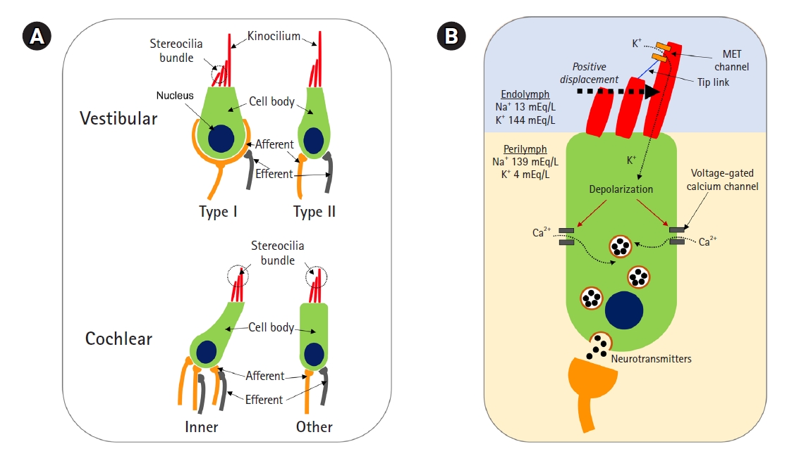
In the inner ear, there are four types of hair cells (Fig. 1A): inner hair cells (IHCs) and outer hair cells (OHCs) in the cochlea and type I and II hair cells in the vestibular system. Each has slight differences in its structure and function. The potassium current is the key to the function of those hair cells. For example, in the IHC, the mechanical movement of lymphatic fluid deflects the stereocilia, opening the mechanoelectrical transduction (MET) channel located in the stereocilia. The MET channel then permits potassium influx, and the IHC is depolarized (Fig. 1B) [4,5]. When the IHC is depolarized, the voltage-gated calcium channel is opened. Next, neurotransmitters are secreted, and auditory nerve fibers are activated [5,6].
Disorders related to inner ear dysfunction, including hearing loss, are mostly caused by damaged hair cells [7]. Of particular note, in mammals, the damage is permanent; hair cells neither regenerate nor recover except in very limited situations. Therefore, research on the inner ear focuses on those hair cells, including the prevention of cell damage and cell regeneration.

2. Research on hearing loss and vestibular disorders

Researchers in this field are more interested in cochlear hair cells (IHCs and OHCs) than in vestibular hair cells (type I and type II hair cells). This is because patients with disequilibrium can recover their balance functionality through vestibular compensation. In fact, balance involves integrating information from vision, proprioception, and the vestibular system. If one function (e.g., the vestibular system) is weakened, the others (e.g., vision and proprioception) are strengthened; as a result, the function of balance can be clinically restored by comprehensive vestibular rehabilitation therapy, allowing a patient to maintain a normal life [8-10].
In contrast, hearing loss resulting from damage to cochlear hair cells (IHCs and OHCs) does not recover except in very few cases. Thus, the prevalence of hearing loss is high; it is estimated to be 15% in all ages and reaches two-thirds in the older (≥70 years) population [11,12]. Furthermore, there is no medical treatment other than using devices, such as hearing aids and cochlear implants (CIs). Thus, in response to a high demand for treatment, research that ultimately aims at the development of drugs targeting hearing loss is actively underway worldwide [13]. Otic organoids are attracting attention because they can be used both as a platform for such research and as cell therapy to promote hair cell regeneration.

3. The organ of Corti

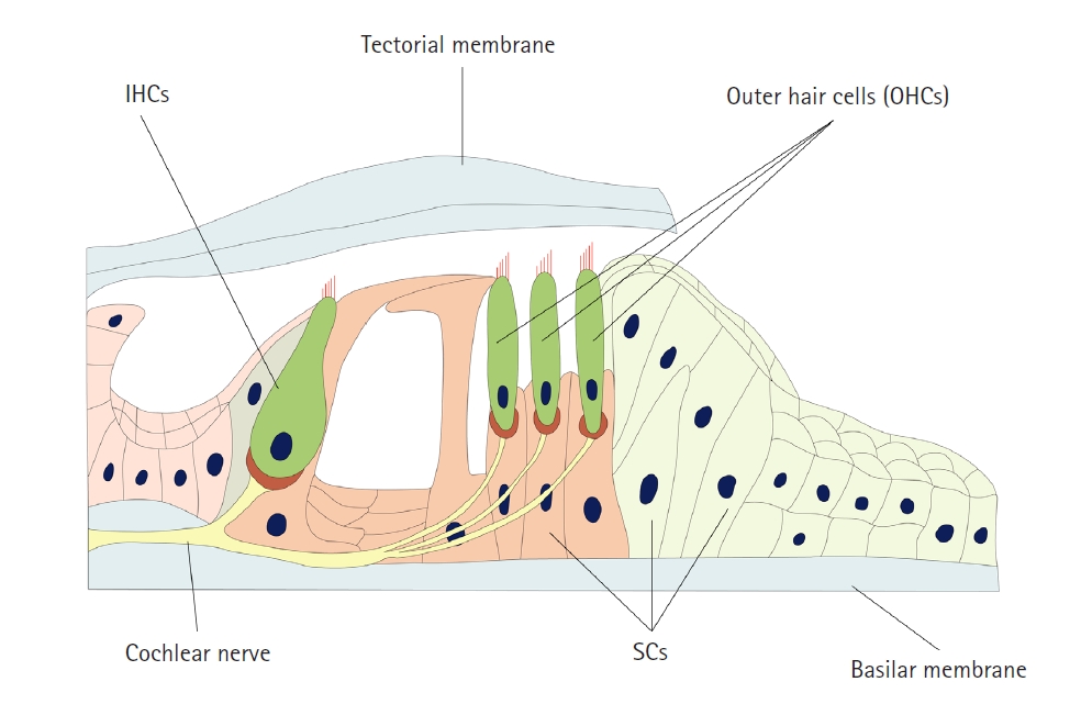
The cochlea is the organ responsible for auditory function. The cochlea is made up of IHCs, OHCs, supporting cells, the tectorial membrane, the basilar membrane, and the spiral ganglion, and those components maintain a structure called the organ of Corti (Fig. 2). This structure—the organ of Corti—plays a key role in hair cell function. The stereocilia of hair cells are contained in the endolymph, which maintains a high concentration of potassium. These cells can be depolarized by the influx of potassium through MET channels. Hair cells synapse with the spiral ganglion, and finally, auditory information is transmitted from the organ of Corti to the brain via the spiral ganglion [5].

4. Otic organoids

To summarize, the key elements in the production of otic organoids are as follows: (1) Differentiation of hair cell-like cells with structural similarities, including stereocilia; (2) Maturation of hair cell-like cells with functional similarities, including ion channels capable of potassium influx; (3) Overall, differentiation and maturation into cochlear hair cells, not vestibular hair cells; (4) Materialization of the organ of Corti, including hair cells, supporting cells, and the extracellular fluid with a high concentration of potassium.
Based on the above issues, this review article will discuss the development and progress of otic organoids, including future directions of research.

Development of otic organoids

Ethics statement: This study was a literature review of previously published studies and was therefore exempt from institutional review board approval.

1. Hair cell regeneration: transdifferentiation from supporting cells and differentiation from pluripotent stem cells

Researchers are interested in hair cell regeneration because most cases of hearing loss are caused by the permanent loss of hair cells. While hair cells cannot be regenerated in mammals, birds and amphibians can regenerate hair cells through two mechanisms: supporting cells may be transformed into hair cells, or they can divide to produce hair cell progenitors [14-16]. Therefore, extensive research has sought to determine how to transdifferentiate supporting cells into hair cells in mammals, and it is still actively underway [17-20].
Hair cell regeneration via transdifferentiation is expected to develop further in the future; however, it also has limitations. There are restrictions in terms of cell therapy that transplants those regenerated cells. For instance, gathering inner ear tissue from patients is not practically feasible, making it impossible to regenerate hair cells containing the genetic information of the patient via transdifferentiation.
Naturally, researchers have studied hair cell differentiation from pluripotent stem cells (PSCs). Initial experiments were based on a two-dimensional (2D) culture system used to generate retinal cells from PSCs [21]. Studies have demonstrated that hair cell-like cells with characteristics of hair cells could be differentiated from mouse and human PSCs [22-27]. However, since the above studies conducted 2D culture in Petri dishes and mainly focused on hair cells only, those cannot be considered as otic organoids.

2. Three-dimensional otic organoids

In 2013, Koehler et al. [28] reported that inner ear sensory epithelia could be differentiated from mouse PSCs using three-dimensional (3D) culture. The inner ear epithelia showed a specific structure similar to the inner ear, where hair cells and supporting cells maintain their own arrangement; these were referred to as the first otic organoids. Furthermore, in 2017, the same research team succeeded in differentiating otic organoids from human PSCs [29]. These researchers are the most advanced group in the development of otic organoids, and the protocols they presented are widely used in the generation of either mouse or human otic organoids [30,31].
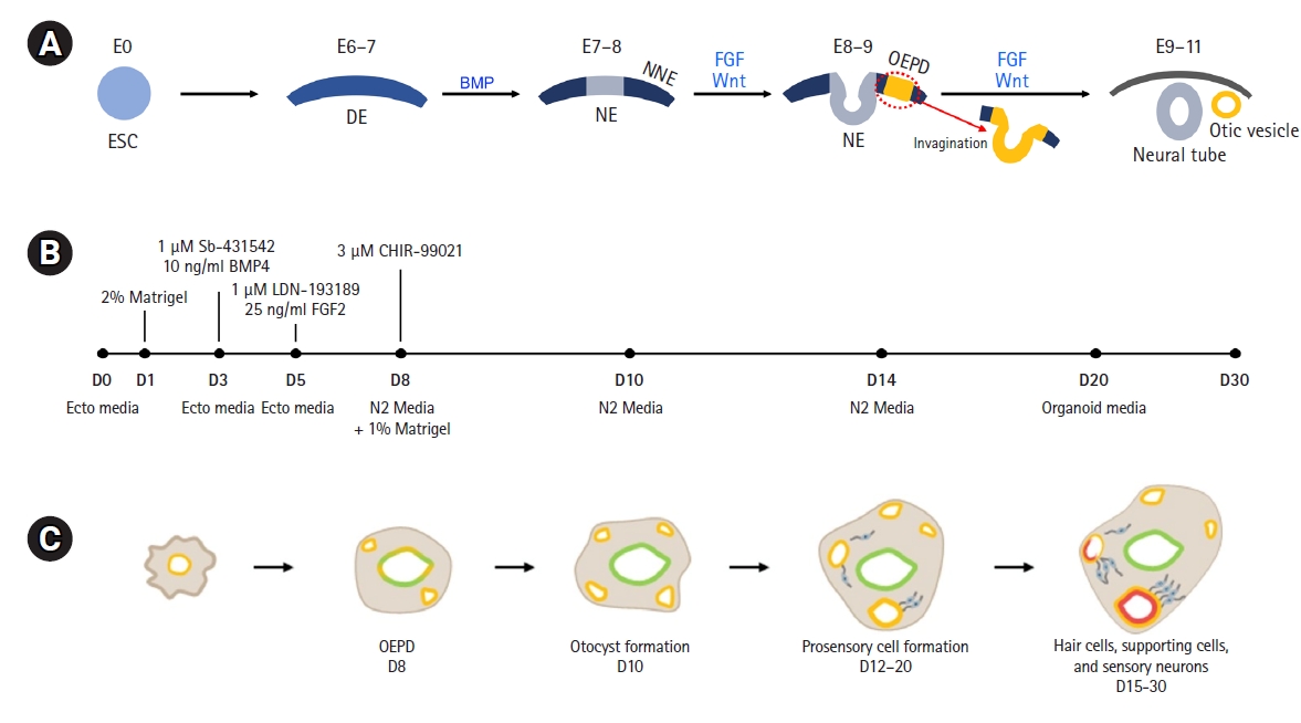
The aim of these protocols is to replicate the intricate and complex natural development process of the inner ear. Prior to a summary of the protocols, we provide a concise overview of the inner ear’s natural development process. The inner ear is derived from the ectoderm. It proceeds through the definite ectoderm (mouse embryonic day [E], 6-7), non-neural ectoderm (NNE) (E7-8), and preplacodal ectoderm [E8], and then the otic-epibranchial placode domain (OEPD) (E8-9) is formed, entering the otic fate. The OEPD invaginates from the surface of the ectoderm to create the otic pit, and then it breaks away from the ectodermal layer to form the otic vesicle (or otocyst). In this process of inner ear development, bone morphogenetic proteins (BMPs), fibroblast growth factors (FGFs), and Wnt signaling are known to play key roles (Fig. 3A) [32,33]. The protocol developed by Koehler et al. [28,30] follows the differentiation process of the inner ear, treating cells with these signal regulators to induce the formation of otic vesicles.
A brief summary of the generation of otic organoids is as follows. Using a 3D floating culture with 2% Matrigel as the extracellular matrix, the first step is to form PSC aggregates in a U-shaped-bottom microplate. Each U-shaped well has 3,000 to 5,000 PSCs. BMP4 for differentiation into NNE and a transforming growth factor-beta (TGF-β) inhibitor (SB-431542) for preventing mesoderm differentiation are administered on in vitro days 3 to 4. This step is critical in distinguishing the generation of otic organoids from other organoids related to the central nervous system. BMPs play a vital role in promoting differentiation into NNE, while inhibiting differentiation into neural ectoderm destined for the central nervous system [30]. On day 5, FGF2 and a BMP inhibitor (LDN-193189) are added to the medium; FGF2 plays a role in driving it towards the OEPD, whereas the BMP inhibitor prevents it from becoming epidermis. On day 8, a Wnt agonist (CHIR-99021) is administered to accelerate otic vesicle formation (Fig. 3B) [28-30]. An otic organoid containing hair cells, supporting cells, and sensory neurons forms in the vesicle. Mouse PSCs take 20 to 30 days for organoid formation, while up to 90 days are required for human PSCs (Fig. 3C).
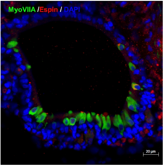
Starting on approximately day 10, these vesicles begin to form around the outer layer of a cell aggregate (Fig. 4). For the next 10 days, the sensory epithelia develop inside the vesicle. From day 20, the alignment of hair cell-like cells and supporting cell-like cells is identified in immunohistochemical staining (Fig. 5). Fig. 5 shows the most typical form of an otic organoid. Some of the immunohistochemical markers utilized for the characterization of cells in otic organoids include Sox2 (for the sensory epithelium including hair cells and supporting cells), MyoVIIa (for the cytosol of hair cells), Brn3c (for the nuclei of hair cells), α-tubulin (for kinocilia), Espin (for stereocilia), βIII-tubulin (for neurons), synaptophysin (for synapses), and Ctbp2 (for ribbon synapses) (for a comprehensive list, refer to Table 1 in the publication by Koehler and Hashino, 2014) [30]. Hair cell-like cells also have stereocilia. In addition, domains related to the synapse with nerves are observed. Although they are somewhat immature, it has been identified that the stereocilia of those hair cell-like cells are not only morphologically differentiated, but also function as ion channels.
Despite significant progress in the development of otic organoids, much remains to be elucidated regarding the incidental structures that may arise during this process. While nerve fiber-like structures have been frequently observed during otic organoid development, their significance beyond their synapse with the organoids remains unclear. Notably, a study has reported the spontaneous development of cardiac structures in some instances, which generate a bearing rhythm. It has been suggested that Wnt modulation may play a role in the formation of these unnecessary structures [34]. These findings underscore the need for continued refinement and improvement of otic organoid development protocols.

Applications of otic organoids in inner ear research

1. Current research platform in inner ear research

To date, hearing loss studies have used in vitro cell lines or in vivo animal models. For in vitro studies, a variety of immortalized cell lines have been developed, which have the characteristics of hair cells [35-37]. Among them, the HEI-OC1 cell line is well-known and is used as a screening tool before animal studies. Although novel research tools have been developed, the HEI-OC1 cell line continues to be widely used. However, we cannot say that HEI-OC1 cells exactly replicate any living beings. In addition, the hair cells are immature, with neither stereocilia nor MET channels [38].
The larval zebrafish lateral line is also used. It was first developed in 2005, and its hair cells are labeled with fluorescence [39]. Zebrafish are employed in studies elucidating the development of hair cells and the mechanisms of diseases, as well as for screening drug compounds [40,41]. The zebrafish lateral line is in vivo, making it superior to experiments using cell lines. However, all research should ultimately be applicable to humans, and the substantial inter-species differences between fish and mammals are the largest hurdle.
Therefore, animal studies still play a role in inner ear research. In addition to administering drugs directly to animals, we can also obtain cochlear explants from neonatal rodents; this method is referred to as ex vivo [42,43]. However, experiments using animals are increasingly facing skepticism; there are not only ethical issues, but also doubts about the effectiveness of the experiments [44,45]. Needless to say, a human experiment would be even more difficult to conduct.

2. Otic organoids as a research platform

Table 1 summarizes the characteristics of research methodologies. Experimentation using organoids may overcome the limitations of each method. Organoids would allow large-number studies because many organoids can be produced simultaneously. In addition, organoids generated from human PSCs naturally have a high similarity to humans. Another major advantage of this method is that it is free from ethical issues.
Due to the inability to biopsy the human inner ear without causing irreversible damage to hearing or balance, the study of human inner ear tissues has been restricted to scarce fetal and cadaveric tissue. Consequently, it has not been feasible to directly culture, infect, or treat inner ear tissue with toxic drugs, unlike other organs. However, the iPSC reprogramming and otic organoid generation technologies provide an exciting opportunity to study cultured tissue with the patient’s genetic information. This technology has enabled more extensive research on hereditary deafness and has already produced meaningful results [46]. A study on hearing loss caused by Tmprss3 mutations utilized otic organoids to elucidate the mechanism underlying the relationship between hearing loss and the mutation [47].
Otic organoids hold potential for investigating inner ear diseases that arise from infections. By directly infecting organoids with viruses or bacteria, the resultant effects can be studied. For example, a study infected otic organoids with COVID-19 to investigate its impact [48]. Additionally, organoids can serve as a platform for researching drug-induced ototoxicity, and the application of otic organoids has already commenced [49].

3. Otic organoids as cell therapy

Since there is no medical therapy for hearing loss, we cannot help but look forward to the therapeutic potential of otic organoids. In particular, there is some expectation that cell therapy might be possible; that is, that hair cells from organoids may replace the damaged cells of a patient. In fact, similar attempts have been made even before the generation of otic organoids. Mouse embryonic stem cells have been injected directly into the inner ear of an animal to determine whether those cells would differentiate into hair cells [50-55]. However, most cells could not be positioned appropriately. Although some cells could find the location—the endolymph space—they differentiated into neither hair cells nor neurons [50,52,53].
Some researchers attempted to transplant more differentiated cells, otic progenitor cells, into the inner ear [56-61]. The results were slightly better than when embryonic stem cells were transplanted; it was identified that those transplanted cells had synaptic connectivity with neurons. However, that finding did not provide sufficient support to attempt this transplantation for therapeutic purposes in humans. To sum up those attempts of cell therapy, at least, transplantation using a more differentiated cell state has a lower risk of teratoma formation and the cells are more likely to attach to the endolymph space [27]. Therefore, better results would be expected when highly differentiated cells from organoids are transplanted.

Current hurdles and future directions of otic organoids

As discussed above, otic organoids may lead to meaningful innovations in inner ear research, and significant progress has already been made. However, there remain many hurdles to overcome.

1. Differentiation into cochlear hair cells

Research on vestibular disorders is, of course, valuable and promising; however, in fact, researchers, clinicians, and patients have much higher expectations for the use of organoids in hearing loss studies. The cause of hearing loss is the loss of cochlear hair cells (IHCs or OHCs). Therefore, the production of otic organoids containing cochlear hair cells would be undoubtedly important for research on hearing loss. However, the properties of hair cells in otic organoids to date are closer to those of vestibular hair cells [28,29,62-64].
In a study using human PSCs, prestin, an OHC marker, was once identified, but except for that study [65], the types of hair cells were vestibular. Considering the inner ear developmental process, the default direction of differentiation after the otic vesicle is the vestibular system. The protocols to date do not contain any additional factors after vesicle formation, so this might be the natural outcome. This fact suggests that the otic organoids that have been generated to date are still immature. Although several efforts have been made for the maturation of otic organoids, a protocol for cochlear hair cell differentiation has not yet been established [34,67-69].
The protocol presented in Fig. 3 aims to promote differentiation up to the otocyst stage, but the actual process of inner ear development requires several important steps even after otocyst formation. Specific patterns of gene expression play a vital role in determining the subsequent stages of development, with two well-known types of patterning called the anterior-posterior (AP) axis and the dorsal-ventral (DV) axis [70]. Once these patterns are established, the development of specific inner ear components becomes possible. According to the AP axis, the neurosensory epithelium develops in the anterior domain of the otocyst while the non-sensory epithelium develops in the posterior domain, with retinoic acid and FGF8 playing essential roles in generating this pattern [71,72]. The DV patterning then determines whether the otocyst develops into the vestibular labyrinth (dorsal) or cochlea (ventral) [33]. Sonic hedgehog (SHH) is known to play a key role in this DV pattern, showing a gradient in which the concentration of SHH constantly increases from the dorsal to the ventral sides (Fig. 6) [73-75]. The current protocol for generating otic organoids does not utilize the signaling mediators mentioned above. To produce more developed otic organoids, it may be necessary to incorporate these additional regulators. However, the process may be complex, as it requires creating a gradient of the signal concentration.
Furthermore, in the process of its development, the inner ear must interact with the central nervous system. For example, SHH is secreted from the neural tube. However, the protocol we have used does not take into account this interaction with the surrounding structures. Therefore, the organoid proceeds toward the vestibular fate, which is the default, and it seems to be immature. As a result, it could be worthwhile to apply a co-culture system that can enable interactions between structures (Fig. 6) [76,77].

2. Yield efficiency and reproducibility in the generation of otic organoids

According to reports, otic organoids were formed in approximately 70% to 80% of all aggregates during differentiation with mouse PSCs, while the formation rate drops to around 20% when using human PSCs [29,64]. It is anticipated that this efficiency will improve over time, but accurately quantifying this efficiency remains challenging. Moreover, there is currently no agreement on how to evaluate the qualitative efficiency of otic organoids. Researchers are completely responsible for manually finding otic vesicles in an aggregate; without immunohistochemistry, it is not known whether a vesicle has been well differentiated. Identified organoids are not consistent in either the number or arrangement of cells.
In addition, the differentiation period is also a hurdle. Using mouse PSCs, an organoid could be completely formed within approximately 30 days, while the process takes 90 days with human PSCs. It is never easy to conduct organoid culture for 90 days without any unexpected problems in the laboratory. This efficiency issue should be solved for the industrialization of otic organoids. The current status might be disappointing, but this means that we should continue making progress until otic organoids can be used as a solid research methodology.

3. Inner ear organoid-on-a-chip

In the generation of otic organoids, it is worth considering the application of a microfluidic-based approach. The organ of Corti, a core structure of the cochlea, has not been developed. Using sophisticated 3D bioprinting to generate an inner ear organoid-on-a-chip, the organ of Corti may be simulated and the signaling gradient (e.g., the concentration gradient of SHH) can be implemented by controlling microfluids on the chip [78]. Investigations on microfluidic or microwell systems are being actively conducted, and these methods are expected to be utilized soon in otic organoids [79-81].

4. Otic organoids versus cochlear implantation

The auditory system in humans is the only sensory organ in our body, the function of which can be restored to the extent that our daily lives are possible via an implantable device—a CI. Cochlear hair cells convert mechanical energy from external and middle ear into electrochemical energy, and that is also what a CI does. Since CIs were first introduced in the 1980s, their effectiveness and safety have been fully demonstrated, and it has been confirmed that CIs can also relieve the social isolation caused by severe hearing loss; thus, the indications for CI surgery are gradually expanding [82-89]. According to the authors’ clinical experience of performing CI surgery and the studies we have conducted, there is no question about the effectiveness of CIs [90-95].
However, CIs cannot be a universal treatment applicable to all hearing loss patients because they require surgery and are indicated for severe hearing loss. The demand for drug treatment for hearing loss is still substantial. Furthermore, surgical experience with CIs will eventually be a major asset for organoid-based cell therapy. Because the electrode of a CI is inserted directly into the inner ear, the same approach can be used as a drug delivery modality (Fig. 7) [96-100]. This means that rapid progress might be possible in the field of otic organoid-based cell therapy.

NOTES

Conflict of interest

No potential conflict of interest relevant to this article was reported.

Funding

This research was supported by Korean Fund for Regenerative Medicine funded by the Ministry of Science and ICT, and Ministry of Health and Welfare (RS-2022-00070599) and supported by the Bio & Medical Technology Development Program of the NRF funded by the Korean government (MSIT) (No. 2019M3A9H1103737).

Author contributions

Conceptualization: HK, JK, KK, YHC; Methodology: HK, YSK, YJK, JH, SS, JHJ, SP, KK; Data curation: HK, YSK, YJK; Visualization: HK, YJK, SP; Project administration: YHC; Supervision: YHC; Writing-original draft: HK, YHC; Writing-review & editing: all authors.

Fig. 1.
(A) Four types of hair cells in the inner ear. As well as their morphological differences, each type has its own function. (B) Positive displacement of stereocilia opens mechanoelectrical transduction (MET) channels mechanically through the tip link, allowing a potassium influx. Then, the cell is depolarized and opens voltage-gated calcium channels. After that, neurotransmitters are secreted.
organoid-2023-3-e7f1.jpg
Fig. 2.
The organ of Corti. Three rows of outer hair cells (OHCs) and one row of inner hair cells (IHCs) are surrounded by supporting cells (SCs). Those cells are located on the basilar membrane. Endolymph in the scala media maintains a high concentration of potassium and a low concentration of sodium (K+ 144 mEq/L and Na+ 13 mEq/L). The movement of the tectorial membrane either opens or closes the mechanoelectrical transduction channel located in the stereocilia of hair cells.
organoid-2023-3-e7f2.jpg
Fig. 3.
(A) Schematic summary of otic development from definite ectoderm (DE) to otocysts in mice. Bone morphogenic proteins (BMPs) are key molecules that induce non-neural ectoderm (NNE). Fibroblast growth factors (FGFs) and Wnt signaling play major roles in the formation of the oto-epibranchial placode domain (OEPD) and otocysts. (B) A protocol for the generation of mouse otic organoids follows the inner ear development from embryonic stem cells (ESCs) to the otocyst. As the extracellular matrix, 2% Matrigel is used. BMP4, FGF2, and Wnt (CHIR-99021) are introduced to induce the otic fate. Transforming growth factor (TGF)-β (SB-431542) inhibits the differentiation from DE to mesoderm. A BMP inhibitor (LDN-193189) prevents differentiation from NNE to epidermis. (C) Schematic figures from cell aggregates to otic organoids. An otocyst (yellow circle) develops into an otic organoid including sensory epithelia (red circle). The organoid has a structure containing hair cells, supporting cells, and sensory neurons. D, day.
organoid-2023-3-e7f3.jpg
Fig. 4.
Microscopic images from a stem cell aggregate to an otic vesicle. (A) The outer layer of a cell aggregate is thickened, differentiating to the oto-epibranchial placode domain (OEPD) (scale bar=100 μm). (B) After day 10, circular vesicles appear, and those become otic organoids (scale bar=200 μm). Differential interference contrast images. Mouse embryonic stem cells (J1 cells; 3,000 cells/well).
organoid-2023-3-e7f4.jpg
Fig. 5.
Immunohistochemistry images of otic organoids. Hair cell-like cells are expressed as MyoVIIa. They show stereocilia marked with Espin (days 20-30).
organoid-2023-3-e7f5.jpg
Fig. 6.
Otic organoids replicating the vestibular system. It is thought that additional stimulations, such as more signaling molecules and interaction with adjacent structures, are necessary to differentiate into the cochlear system. ① Signaling molecules may need to be implemented in a gradient. ② Co-culture may be required to replicate the interactions with adjacent structures.
organoid-2023-3-e7f6.jpg
Fig. 7.
Drug elution through a cochlear implant device. An electrode array is directly inserted into the cochlea. It can be located very close to the organ of Corti, which is the target for cell therapy.
organoid-2023-3-e7f7.jpg
Table 1.
Summary of methodologies in inner ear research
Cell lines Zebrafish Organ of Corti explantation Mammalian animal studies Human studies
Large-number studies Possible Possible Impossible Impossible Impossible
Disease modeling Partially possible Possible Possible Possible Impossible
Similarity to humans None Very low Low Low Very high
Ethical issues None Low High High Very high

References

1. Torres M, Giráldez F. The development of the vertebrate inner ear. Mech Dev 1998;71:5-21.
crossref pmid
2. Fettiplace R. Hair cell transduction, tuning, and synaptic transmission in the mammalian cochlea. Compr Physiol 2017;7:1197-227.
crossref pmid pmc pdf
3. Hudspeth AJ. How the ear’s works work: mechanoelectrical transduction and amplification by hair cells. C R Biol 2005;328:155-62.
crossref pmid
4. Beurg M, Fettiplace R, Nam JH, Ricci AJ. Localization of inner hair cell mechanotransducer channels using high-speed calcium imaging. Nat Neurosci 2009;12:553-8.
crossref pmid pmc pdf
5. Schwander M, Kachar B, Müller U. Review series: the cell biology of hearing. J Cell Biol 2010;190:9-20.
pmid pmc
6. Hudspeth AJ, Lewis RS. A model for electrical resonance and frequency tuning in saccular hair cells of the bull-frog, Rana catesbeiana. J Physiol 1988;400:275-97.
crossref pmid pmc
7. Wagner EL, Shin JB. Mechanisms of hair cell damage and repair. Trends Neurosci 2019;42:414-24.
crossref pmid pmc
8. Helmchen C, Klinkenstein JC, Krüger A, Gliemroth J, Mohr C, Sander T. Structural brain changes following peripheral vestibulo-cochlear lesion may indicate multisensory compensation. J Neurol Neurosurg Psychiatry 2011;82:309-16.
crossref pmid
9. Parietti-Winkler C, Gauchard GC, Simon C, Perrin PP. Long-term effects of vestibular compensation on balance control and sensory organisation after unilateral deafferentation due to vestibular schwannoma surgery. J Neurol Neurosurg Psychiatry 2010;81:934-6.
crossref pmid
10. zu Eulenburg P, Stoeter P, Dieterich M. Voxel-based morphometry depicts central compensation after vestibular neuritis. Ann Neurol 2010;68:241-9.
crossref pmid
11. Mahboubi H, Lin HW, Bhattacharyya N. Prevalence, characteristics, and treatment patterns of hearing difficulty in the United States. JAMA Otolaryngol Head Neck Surg 2018;144:65-70.
crossref pmid
12. Lin FR, Thorpe R, Gordon-Salant S, Ferrucci L. Hearing loss prevalence and risk factors among older adults in the United States. J Gerontol A Biol Sci Med Sci 2011;66:582-90.
crossref pmid
13. Müller U, Barr-Gillespie PG. New treatment options for hearing loss. Nat Rev Drug Discov 2015;14:346-65.
crossref pmid pdf
14. Lewis RM, Keller JJ, Wan L, Stone JS. Bone morphogenetic protein 4 antagonizes hair cell regeneration in the avian auditory epithelium. Hear Res 2018;364:1-11.
crossref pmid pmc
15. Matsunaga M, Kita T, Yamamoto R, Yamamoto N, Okano T, Omori K, et al. Initiation of supporting cell activation for hair cell regeneration in the avian auditory epithelium: an explant culture model. Front Cell Neurosci 2020;14:583994.
crossref pmid pmc
16. Stone JS, Cotanche DA. Hair cell regeneration in the avian auditory epithelium. Int J Dev Biol 2007;51:633-47.
crossref pmid
17. Franco B, Malgrange B. Concise review: regeneration in mammalian cochlea hair cells: help from supporting cells transdifferentiation. Stem Cells 2017;35:551-6.
crossref pmid pdf
18. Yang J, Cong N, Han Z, Huang Y, Chi F. Ectopic hair cell-like cell induction by Math1 mainly involves direct transdifferentiation in neonatal mammalian cochlea. Neurosci Lett 2013;549:7-11.
crossref pmid
19. Defourny J, Mateo Sánchez S, Schoonaert L, Robberecht W, Davy A, Nguyen L, et al. Cochlear supporting cell transdifferentiation and integration into hair cell layers by inhibition of ephrin-B2 signalling. Nat Commun 2015;6:7017.
crossref pmid pdf
20. Shu Y, Li W, Huang M, Quan YZ, Scheffer D, Tian C, et al. Renewed proliferation in adult mouse cochlea and regeneration of hair cells. Nat Commun 2019;10:5530.
crossref pmid pmc pdf
21. Lamba DA, Karl MO, Ware CB, Reh TA. Efficient generation of retinal progenitor cells from human embryonic stem cells. Proc Natl Acad Sci U S A 2006;103:12769-74.
crossref pmid pmc
22. Oshima K, Shin K, Diensthuber M, Peng AW, Ricci AJ, Heller S. Mechanosensitive hair cell-like cells from embryonic and induced pluripotent stem cells. Cell 2010;141:704-16.
crossref pmid pmc
23. Ouji Y, Ishizaka S, Nakamura-Uchiyama F, Yoshikawa M. In vitro differentiation of mouse embryonic stem cells into inner ear hair cell-like cells using stromal cell conditioned medium. Cell Death Dis 2012;3:e314.
crossref pmid pmc pdf
24. Ohnishi H, Skerleva D, Kitajiri S, Sakamoto T, Yamamoto N, Ito J, et al. Limited hair cell induction from human induced pluripotent stem cells using a simple stepwise method. Neurosci Lett 2015;599:49-54.
crossref pmid
25. Ronaghi M, Nasr M, Ealy M, Durruthy-Durruthy R, Waldhaus J, Diaz GH, et al. Inner ear hair cell-like cells from human embryonic stem cells. Stem Cells Dev 2014;23:1275-84.
crossref pmid pmc
26. Ding J, Tang Z, Chen J, Shi H, Chen J, Wang C, et al. Induction of differentiation of human embryonic stem cells into functional hair-cell-like cells in the absence of stromal cells. Int J Biochem Cell Biol 2016;81(Pt A):208-22.
crossref pmid
27. Tang PC, Hashino E, Nelson RF. Progress in modeling and targeting inner ear disorders with pluripotent stem cells. Stem Cell Reports 2020;14:996-1008.
crossref pmid pmc
28. Koehler KR, Mikosz AM, Molosh AI, Patel D, Hashino E. Generation of inner ear sensory epithelia from pluripotent stem cells in 3D culture. Nature 2013;500:217-21.
crossref pmid pmc pdf
29. Koehler KR, Nie J, Longworth-Mills E, Liu XP, Lee J, Holt JR, et al. Generation of inner ear organoids containing functional hair cells from human pluripotent stem cells. Nat Biotechnol 2017;35:583-9.
crossref pmid pmc pdf
30. Koehler KR, Hashino E. 3D mouse embryonic stem cell culture for generating inner ear organoids. Nat Protoc 2014;9:1229-44.
crossref pmid pmc pdf
31. Nie J, Hashino E. Generation of inner ear organoids from human pluripotent stem cells. Methods Cell Biol 2020;159:303-21.
crossref pmid
32. Sai X, Ladher RK. Early steps in inner ear development: induction and morphogenesis of the otic placode. Front Pharmacol 2015;6:19.
crossref pmid pmc
33. Whitfield TT. Development of the inner ear. Curr Opin Genet Dev 2015;32:112-8.
crossref pmid
34. Carpena NT, Chang SY, Choi JE, Jung JY, Lee MY. Wnt modulation enhances otic differentiation by facilitating the enucleation process but develops unnecessary cardiac structures. Int J Mol Sci 2021;22:10306.
crossref pmid pmc
35. Barald KF, Lindberg KH, Hardiman K, Kavka AI, Lewis JE, Victor JC, et al. Immortalized cell lines from embryonic avian and murine otocysts: tools for molecular studies of the developing inner ear. Int J Dev Neurosci 1997;15:523-40.
crossref pmid pdf
36. Holley MC, Lawlor PW. Production of conditionally immortalised cell lines from a transgenic mouse. Audiol Neurootol 1997;2:25-35.
crossref pmid
37. Kalinec F, Kalinec G, Boukhvalova M, Kachar B. Establishment and characterization of conditionally immortalized organ of corti cell lines. Cell Biol Int 1999;23:175-84.
crossref pmid
38. Kalinec GM, Webster P, Lim DJ, Kalinec F. A cochlear cell line as an in vitro system for drug ototoxicity screening. Audiol Neurootol 2003;8:177-89.
crossref pmid pdf
39. Ton C, Parng C. The use of zebrafish for assessing ototoxic and otoprotective agents. Hear Res 2005;208:79-88.
crossref pmid
40. Owens KN, Santos F, Roberts B, Linbo T, Coffin AB, Knisely AJ, et al. Identification of genetic and chemical modulators of zebrafish mechanosensory hair cell death. PLoS Genet 2008;4:e1000020.
crossref pmid pmc
41. Coffin AB, Williamson KL, Mamiya A, Raible DW, Rubel EW. Profiling drug-induced cell death pathways in the zebrafish lateral line. Apoptosis 2013;18:393-408.
crossref pmid pmc pdf
42. Low W, Dazert S, Baird A, Ryan AF. Basic fibroblast growth factor (FGF-2) protects rat cochlear hair cells in organotypical culture from aminoglycoside injury. J Cell Physiol 1996;167:443-50.
crossref pmid
43. Corbacella E, Lanzoni I, Ding D, Previati M, Salvi R. Minocycline attenuates gentamicin induced hair cell loss in neonatal cochlear cultures. Hear Res 2004;197:11-8.
crossref pmid
44. Pound P, Ritskes-Hoitinga M. Is it possible to overcome issues of external validity in preclinical animal research?: why most animal models are bound to fail. J Transl Med 2018;16:304.
crossref pmid pmc pdf
45. Akhtar A. The flaws and human harms of animal experimentation. Camb Q Healthc Ethics 2015;24:407-19.
crossref pmid pmc
46. Romano DR, Hashino E, Nelson RF. Deafness-in-a-dish: modeling hereditary deafness with inner ear organoids. Hum Genet 2022;141:347-62.
crossref pmid pdf
47. Tang PC, Alex AL, Nie J, Lee J, Roth AA, Booth KT, et al. Defective Tmprss3-associated hair cell degeneration in inner ear organoids. Stem Cell Reports 2019;13:147-62.
crossref pmid pmc
48. Jeong M, Ocwieja KE, Han D, Wackym PA, Zhang Y, Brown A, et al. Direct SARS-CoV-2 infection of the human inner ear may underlie COVID-19-associated audiovestibular dysfunction. Commun Med (Lond) 2021;1:44.
crossref pmid pmc pdf
49. Kurihara S, Fujioka M, Hirabayashi M, Yoshida T, Hosoya M, Nagase M, et al. Otic organoids containing spiral ganglion neuron-like cells derived from human-induced pluripotent stem cells as a model of drug-induced neuropathy. Stem Cells Transl Med 2022;11:282-96.
crossref pmid pmc pdf
50. Sakamoto T, Nakagawa T, Endo T, Kim TS, Iguchi F, Naito Y, et al. Fates of mouse embryonic stem cells transplanted into the inner ears of adult mice and embryonic chickens. Acta Otolaryngol Suppl 2004;(551):48-52.
crossref
51. Hu Z, Ulfendahl M, Olivius NP. Central migration of neuronal tissue and embryonic stem cells following transplantation along the adult auditory nerve. Brain Res 2004;1026:68-73.
crossref pmid
52. Regala C, Duan M, Zou J, Salminen M, Olivius P. Xenografted fetal dorsal root ganglion, embryonic stem cell and adult neural stem cell survival following implantation into the adult vestibulocochlear nerve. Exp Neurol 2005;193:326-33.
crossref pmid
53. Lang H, Schulte BA, Goddard JC, Hedrick M, Schulte JB, Wei L, et al. Transplantation of mouse embryonic stem cells into the cochlea of an auditory-neuropathy animal model: effects of timing after injury. J Assoc Res Otolaryngol 2008;9:225-40.
crossref pmid pmc pdf
54. Praetorius M, Vicario I, Schimmang T. Efficient transfer of embryonic stem cells into the cochlea via a non-invasive vestibular route. Acta Otolaryngol 2008;128:720-3.
crossref pmid
55. Zhao LD, Li L, Wu N, Li DK, Ren LL, Guo WW, et al. Migration and differentiation of mouse embryonic stem cells transplanted into mature cochlea of rats with aminoglycoside-induced hearing loss. Acta Otolaryngol 2013;133:136-43.
crossref pmid
56. Corrales CE, Pan L, Li H, Liberman MC, Heller S, Edge AS. Engraftment and differentiation of embryonic stem cell-derived neural progenitor cells in the cochlear nerve trunk: growth of processes into the organ of Corti. J Neurobiol 2006;66:1489-500.
crossref pmid pmc
57. Okano T, Nakagawa T, Endo T, Kim TS, Kita T, Tamura T, et al. Engraftment of embryonic stem cell-derived neurons into the cochlear modiolus. Neuroreport 2005;16:1919-22.
crossref pmid
58. Coleman B, Hardman J, Coco A, Epp S, de Silva M, Crook J, et al. Fate of embryonic stem cells transplanted into the deafened mammalian cochlea. Cell Transplant 2006;15:369-80.
crossref pmid pdf
59. Matsumoto M, Nakagawa T, Kojima K, Sakamoto T, Fujiyama F, Ito J. Potential of embryonic stem cell-derived neurons for synapse formation with auditory hair cells. J Neurosci Res 2008;86:3075-85.
crossref pmid
60. Chen W, Jongkamonwiwat N, Abbas L, Eshtan SJ, Johnson SL, Kuhn S, et al. Restoration of auditory evoked responses by human ES-cell-derived otic progenitors. Nature 2012;490:278-82.
crossref pmid pmc pdf
61. Nishimura K, Nakagawa T, Sakamoto T, Ito J. Fates of murine pluripotent stem cell-derived neural progenitors following transplantation into mouse cochleae. Cell Transplant 2012;21:763-71.
crossref pmid pdf
62. Lahlou H, Nivet E, Lopez-Juarez A, Fontbonne A, Assou S, Zine A. Enriched differentiation of human otic sensory progenitor cells derived from induced pluripotent stem cells. Front Mol Neurosci 2018;11:452.
crossref pmid pmc
63. Mattei C, Lim R, Drury H, Nasr B, Li Z, Tadros MA, et al. Generation of vestibular tissue-like organoids from human pluripotent stem cells using the rotary cell culture system. Front Cell Dev Biol 2019;7:25.
crossref pmid pmc
64. Liu XP, Koehler KR, Mikosz AM, Hashino E, Holt JR. Functional development of mechanosensitive hair cells in stem cell-derived organoids parallels native vestibular hair cells. Nat Commun 2016;7:11508.
crossref pmid pmc pdf
65. Jeong M, O’Reilly M, Kirkwood NK, Al-Aama J, Lako M, Kros CJ, et al. Generating inner ear organoids containing putative cochlear hair cells from human pluripotent stem cells. Cell Death Dis 2018;9:922.
crossref pmid pmc pdf
66. Moeinvaziri F, Shojaei A, Haghparast N, Yakhkeshi S, Nemati S, Hassani SN, et al. Towards maturation of human otic hair cell-like cells in pluripotent stem cell-derived organoid transplants. Cell Tissue Res 2021;386:321-33.
crossref pmid pdf
67. Hocevar SE, Liu L, Duncan RK. Matrigel is required for efficient differentiation of isolated, stem cell-derived otic vesicles into inner ear organoids. Stem Cell Res 2021;53:102295.
crossref pmid pmc
68. Chang SY, Carpena NT, Mun S, Jung JY, Chung PS, Shim H, et al. Enhanced inner-ear organoid formation from mouse embryonic stem cells by photobiomodulation. Mol Ther Methods Clin Dev 2020;17:556-67.
crossref pmid pmc
69. Chang SY, Lee MY. Photobiomodulation with a wavelength > 800 nm induces morphological changes in stem cells within otic organoids and scala media of the cochlea. Lasers Med Sci 2021;36:1917-25.
crossref pmid pdf
70. Nakajima Y. Signaling regulating inner ear development: cell fate determination, patterning, morphogenesis, and defects. Congenit Anom (Kyoto) 2015;55:17-25.
crossref pmid
71. Bok J, Raft S, Kong KA, Koo SK, Dräger UC, Wu DK. Transient retinoic acid signaling confers anterior-posterior polarity to the inner ear. Proc Natl Acad Sci U S A 2011;108:161-6.
crossref pmid
72. Abelló G, Khatri S, Radosevic M, Scotting PJ, Giráldez F, Alsina B. Independent regulation of Sox3 and Lmx1b by FGF and BMP signaling influences the neurogenic and non-neurogenic domains in the chick otic placode. Dev Biol 2010;339:166-78.
crossref pmid
73. Riccomagno MM, Martinu L, Mulheisen M, Wu DK, Epstein DJ. Specification of the mammalian cochlea is dependent on Sonic hedgehog. Genes Dev 2002;16:2365-78.
crossref pmid pmc
74. Bok J, Bronner-Fraser M, Wu DK. Role of the hindbrain in dorsoventral but not anteroposterior axial specification of the inner ear. Development 2005;132:2115-24.
crossref pmid pdf
75. Dessaud E, McMahon AP, Briscoe J. Pattern formation in the vertebrate neural tube: a sonic hedgehog morphogen-regulated transcriptional network. Development 2008;135:2489-503.
crossref pmid pdf
76. Butts JC, McCreedy DA, Martinez-Vargas JA, Mendoza-Camacho FN, Hookway TA, Gifford CA, et al. Differentiation of V2a interneurons from human pluripotent stem cells. Proc Natl Acad Sci U S A 2017;114:4969-74.
crossref pmid pmc
77. Birey F, Andersen J, Makinson CD, Islam S, Wei W, Huber N, et al. Assembly of functionally integrated human forebrain spheroids. Nature 2017;545:54-9.
crossref pmid pmc pdf
78. van der Valk WH, Steinhart MR, Zhang J, Koehler KR. Building inner ears: recent advances and future challenges for in vitro organoid systems. Cell Death Differ 2021;28:24-34.
crossref pmid pdf
79. Zheng Y, Xue X, Shao Y, Wang S, Esfahani SN, Li Z, et al. Controlled modelling of human epiblast and amnion development using stem cells. Nature 2019;573:421-5.
crossref pmid pmc pdf
80. Manfrin A, Tabata Y, Paquet ER, Vuaridel AR, Rivest FR, Naef F, et al. Engineered signaling centers for the spatially controlled patterning of human pluripotent stem cells. Nat Methods 2019;16:640-8.
crossref pmid pdf
81. Brandenberg N, Hoehnel S, Kuttler F, Homicsko K, Ceroni C, Ringel T, et al. High-throughput automated organoid culture via stem-cell aggregation in microcavity arrays. Nat Biomed Eng 2020;4:863-74.
crossref pmid pdf
82. Carlson ML. Cochlear implantation in adults. N Engl J Med 2020;382:1531-42.
crossref pmid
83. Teagle HF, Park LR, Brown KD, Zdanski C, Pillsbury HC. Pediatric cochlear implantation: a quarter century in review. Cochlear Implants Int 2019;20:288-98.
crossref pmid
84. Yoon PJ. Pediatric cochlear implantation. Curr Opin Pediatr 2011;23:346-50.
crossref pmid
85. Claes AJ, Van de Heyning P, Gilles A, Van Rompaey V, Mertens G. Cognitive outcomes after cochlear implantation in older adults: a systematic review. Cochlear Implants Int 2018;19:239-54.
crossref pmid
86. Blebea CM, Ujvary LP, Necula V, Dindelegan MG, Perde-Schrepler M, Stamate MC, et al. Current concepts and future trends in increasing the benefits of cochlear implantation: a narrative review. Medicina (Kaunas) 2022;58:747.
crossref pmid pmc
87. Hempel JM, Simon F, Müller JM. Extended applications for cochlear implantation. Adv Otorhinolaryngol 2018;81:74-80.
crossref pmid
88. Andries E, Gilles A, Topsakal V, Vanderveken OM, Van de Heyning P, Van Rompaey V, et al. Systematic review of quality of life assessments after cochlear implantation in older adults. Audiol Neurootol 2021;26:61-75.
crossref pmid pdf
89. Gaylor JM, Raman G, Chung M, Lee J, Rao M, Lau J, et al. Cochlear implantation in adults: a systematic review and meta-analysis. JAMA Otolaryngol Head Neck Surg 2013;139:265-72.
crossref pmid
90. Jang JH, Roh JM, Choo OS, Kim YJ, Kim H, Park HY, et al. Critical factors for binaural hearing in children with bilateral sequential cochlear implantation: first implant performance and inter-implant interval. Audiol Neurootol 2019;24:174-82.
crossref pmid pdf
91. Choo OS, Kim H, Kim YJ, Roh J, Jang JH, Park HY, et al. Effect of age at cochlear implantation in educational placement and peer relationships. Ear Hear 2021;42:1054-61.
crossref pmid
92. Kim H, Ha J, Gil ES, Jang JH, Park HY, Choung YH. Selection of the optimal first ear for sequential bilateral cochlear implantation in children. Ear Nose Throat J 2021 Dec 13 [Epub]. https://doi.org/10.1177/01455613211064012.
crossref
93. Jang JH, Choo OS, Kim H, Park HY, Choung YH. Round window membrane visibility related to success of hearing preservation in cochlear implantation. Acta Otolaryngol 2019;139:618-24.
crossref pmid
94. Jang JH, Mun HA, Choo OS, Park HY, Choung YH. The speech perception after cochlear implantation: the hearing gain difference according to the implant systems is important? Auris Nasus Larynx 2019;46:330-4.
crossref pmid
95. Kim H, Choo OS, Ha J, Jang JH, Park HY, Choung YH. A new CT parameter for predicting residual hearing preservation in cochlear implantation: the “basal turn-facial ridge angle”. Otol Neurotol 2021;42:e161-7.
crossref pmid
96. Astolfi L, Simoni E, Giarbini N, Giordano P, Pannella M, Hatzopoulos S, et al. Cochlear implant and inflammation reaction: safety study of a new steroid-eluting electrode. Hear Res 2016;336:44-52.
crossref pmid
97. Dhanasingh A, Hochmair I. Drug delivery in cochlear implantation. Acta Otolaryngol 2021;141(sup1):135-56.
crossref
98. Manrique-Huarte R, Zulueta-Santos C, Calavia D, Linera-Alperi MÁ, Gallego MA, Jolly C, et al. Cochlear implantation with a dexamethasone eluting electrode array: functional and anatomical changes in non-human primates. Otol Neurotol 2020;41:e812-22.
crossref pmid
99. Huang Y, Yu H, Liang M, Hou S, Chen J, Zhang F, et al. Hearing protection outcomes of analog electrode arrays coated with different drug-eluting polymer films implanted into guinea pig cochleae. Drug Des Devel Ther 2021;15:3443-50.
crossref pmid pmc pdf
100. Lee MY, Kim YC, Jang J, Jung JY, Choi H, Jang JH, et al. Dexamethasone delivery for hearing preservation in animal cochlear implant model: continuity, long-term release, and fast release rate. Acta Otolaryngol 2020;140:713-22.
crossref pmid


ABOUT
BROWSE ARTICLES
EDITORIAL POLICY
FOR CONTRIBUTORS
Editorial Office
Room 319, Hall 1 of Chonbuk National University Dental College, 20, Geonji-ro, Deokjin-gu, Jeonju 54907, Korea
Tel: +82-63-270-4024    E-mail: editor@j-organoid.org                

Copyright © 2024 by The Organoid Society.

Developed in M2PI

Close layer
prev next