Organoid > Volume 3; 2023 > Article
Lee, Park, Sung, Lee, and Kim: A polymer-based artificial microenvironment for enhancing cell adhesion

Abstract

Background

The ability to control the cell-surface network is widely used to observe the regulation of host-biomaterial interactions, predict cell behavior, and perform solid organ tissue engineering. We further investigated Nafion from the perspective of cell adhesion and biocompatibility.

Methods

The flexible Nafion micro-patterned mold was fabricated by asilicon master (linewidth, 800 nm; space, 800 nm; depth, 600 nm; line pattern). Four different molds were also fabricated based on PDMS, PUA, acryl, and Teflon. The PUA, acryl, and Teflon molds were exposed under a 365-nm UV lamp for 90 seconds at 40 mJ/s for curing.

Results

We developed micro-pattern poly(tetrafluoroethylene-co-perfluoro-3,6-dioxa-4-meth­yl-7-octene-sulfonic acid) (Nafion) films fabricated by a molding process. We present the fabrication and characterization of flexible, micro-patterned Nafion films and the evaluation of cell adhesion and alignment on these films.

Conclusion

We found that cell adhesion and migration/direction could be modulated by controlling the surface architecture.

Introduction

Dynamic control of cell adhesion to a surface is important for understanding cell networks, their involvement with the extracellular matrix, and the formation of controlled structures for cell-matrix interactions similar to those in native tissues [1-3]. The ability to control the cell-surface network is widely used to observe the regulation of host-biomaterial interactions, predict cell behavior, and perform solid organ tissue engineering [4,5].
Generating thin films as cell adhesion platforms is of great interest because of their applications in biosensor, drug, and delivery research and soft robotics [6]. These platforms should be able to support cell growth while maintaining stability and control cellular ability in order to provide insights into cellular interactions and dynamics, while forming scaffolds and cell-based biosensors [7,8]. In recent research, cellular micro-patterning and organization have been widely used to control cell adhesion onto substrates. Dynamic control of cells has been achieved using surface chemistry based on self-assembled monolayers [9-13], etching [14,15], microfluidic techniques [16,17], photolithography [18,19], and screen printing [20,21]. Surface modifications based on biomolecules, such as enzymes and other proteins, have also been used to enhance cell adhesion [22,23]. Despite their considerable advantages, these methods still suffer from limitations in being applied for flexible and soft substrates because of their rigidity. The Haraguchi group described a flexible substrate with a micro- or nano-pattern that had the ability to enhance cell growth and adhesion, generating ordered and functional cell sheets [22].
Many studies on cell adhesion platforms on flexible, micro- and nano-patterned substrates have reported the use of polystyrene film, which is deposited using spin coating [24], and ultrathin poly(methyl methacrylate) films, which are deposited by screen printing [25]. However, these flexible materials are not biodegradable, limiting their applicability to bio-platforms. Nafion poly(tetrafluoroethylene-co-perfluoro-3,6-dioxa-4-methyl-7-octene-sulfonic acid), a hydrophobic polymer with sulfonic acid groups as ionomeric components, is a material with considerable potential for flexible, nano- or micro-patterned substrates because of its properties. Specifically, Nafion has good thermal and mechanical properties and superior electrical properties as a proton exchange conductor. Furthermore, Nafion-based electrochemical sensors as drug delivery devices have been developed in the biosensor field [26-28].
Herein, we further investigated Nafion from the perspective of cell adhesion and biocompatibility. We demonstrated flexible, strong, micro-patterned Nafion sheets for the adhesion and orientation of cells. Nafion sheets were micro-patterned using an imprinting process, and polydimethylsiloxane (PDMS), polyurethane-acrylate (PUA), teflon, and acryl were used as molded material. Through an optimization process, we demonstrated that acryl showed the best performance. Furthermore, different substrates were used to optimize the process. By modulating the surface architecture of the Nafion micro-pattern, cell adhesion and spreading can be controlled. The fabrication process in this research is simple, rapid, and highly reproducible. Furthermore, the Nafion micro-pattern is separate from the substrate, making it straightforward to form micro-patterns with different mechanical properties. The proposed Nafion micro-pattern can be applied to study cellular interactions by controlling and directing the adherent cells.

Materials and Methods

Ethics statement: The Cell-line used for Cell adhesion test in this study was purchased from commercially avialible vendor, KCLB, and was therefoe exempt form institutional review board approval.

1. Reagents and chemicals

Two commercially available ultraviolet (UV) curable molds (PUA, acryl), one thermal curable mold (PDMS), and one self-made UV curable mold (Teflon) were used. A PUA mold (MINS-311RM; Minuta Technology, Osan, South Korea), an acryl mold (OrmoStamp; Micro Resist Technology GmbH, Berlin, Germany), PDMS (SYLGARD 184 Silicon Elastomer Kit; Dow Corning, Midland, MI, USA), Teflon (Fluolink MD 700; Solvay, Brussels, Belgium; and 1-hydroxycyclohexyl phenyl ketone; Sigma-Aldrich, St. Louis, MO, USA), polyethylene terephthalate (PET) (SH71S; SKC, Seoul, South Korea) were used. In the cell culture process, Dulbecco’s modified Eagle’s Medium (DMEM; Welgene, Gyeongsan, South Korea), fetal bovine serum (Welgene), penicillin-streptomycin (Welgene), glutaraldehyde solution (Sigma-Aldrich), ethanol (Daejung, Busan, South Korea), phosphate-buffered saline (Sigma-Aldrich), and osmium tetroxide solution (Sigma-Aldrich) were used.

2. Micro-patterning of Nafion

The flexible Nafion micro-patterned mold was fabricated by a silicon master (linewidth, 800 nm; space, 800 nm; depth, 600 nm; line pattern). Four different molds were also fabricated based on PDMS, PUA, acryl, and Teflon. The PUA, acryl, and Teflon molds were exposed under a 365-nm UV lamp for 90 s at 40 mJ/s for curing. To create the PDMS mold, a PDMS solution mixed with an elastomer and curing agent (10:1) was dropped onto the PUA mold and incubated for 2 hours at 80°C.
After that, 3 substrates (Si wafer, glass wafer, PET film) were exposed to O2 plasma for 3 minutes at 15 sccm (120 mTorr, 50 W, LF-plasmaster 100; JNE, Korea) to make a hydrophilic surface. Then, a thin film of Nafion was deposited by spin coating (Spin 300 A; Midas System, Daejeon, South Korea) with 5% Nafion (Nafion perfluorinated resin solution; Sigma-Aldrich) solution as the substrate. The micro-pattern was fabricated using a mold by thermal nanoimprinting (T-NIL; Midas System) for 10 minutes at 105°C and 10 bar.

3. Cell culture

The sensors were first sterilized by immersion in 70% ethanol (Daejung), followed by UV light exposure for 20 minutes. Then, MDA-MB-231 (Korean Cell Line Bank; Seoul, South Korea) and MCF-7 (Korean Cell Line Bank) were seeded on each sample in a six-well plate (SPL life Science, Pocheon, Korea) and incubated at 37℃ and 5% CO2 in DMEM supplemented with 10% (v/v) fetal bovine serum, and 1% (v/v) penicillin-streptomycin.

4. Characterization of the Nafion micro-pattern

1) Scanning electron microscopy

Scanning electron microscopy (SEM) micrographs were recorded with a JSM-7610F (JEOL; Tokyo, Japan). The samples were gold-sputtered before the microscopic analyses.

2) Atomic force microscopy

The surface morphology of the gold surfaces was investigated using atomic force microscopy (AFM) (AFM5300E; Hitachi, Tokyo, Japan). A normal tapping mode of the silicon cantilever with an oscillation frequency of 365 kHz and spring constant of 47 N/m (NCH-10V; Digital Instruments, Tonawanda, NY, USA) was used for AFM imaging. No destruction of the sample surface was noticed during imaging. All images are presented in the height mode, where higher parts appear brighter.

Results

1. Fabrication and characterization of micro-patterned Nafion films

Mechanically flexible, biocompatible sheets can be used as platforms for applications of biosensors and tissue scaffolds [29]. A micro-pattern can give an opportunity to guide cells or control the cell morphology. In this work, micro-fabrication of Nafion films was performed using various molds and substrates and patterned by photolithographic techniques. Nafion is suitable for use in cell adhesion because it provides a solid support, expressing a high surface charge density and good water wettability. To make a micro-pattern Nafion film, we chose a Nafion solution, not a Nafion membrane, because the Nafion solution has advantages for making patterns on diverse substrates and electrodes. As previously stated, we used different substrates to optimize the micro-pattern, so we chose the Nafion solution. First, a soft mold was fabricated to make a micro-pattern Nafion film, and we used PDMS, PUA, Teflon, and acryl molds to optimize the micro-pattern of the Nafion film. A soft mold was fabricated using a silicon master, and the micro-pattern was fabricated by thermal lithography using these molds as a base (Fig. 1A). To demonstrate the structural integrity and scalability at the microscale, SEM images were taken. The original dimensions of the pattern were 800 nm linewidth, 800 nm space, and 600 nm height. As shown in Fig. 1B, each mold had diverse dimensions; of particular note, the PDMS mold had reduced linewidth, height, and space. Furthermore, some pattern destruction was observed in the Teflon mold. Of these options, acryl showed the best performance and accurate dimensions (800 nm linewidth, 790 nm space, 590 nm height).
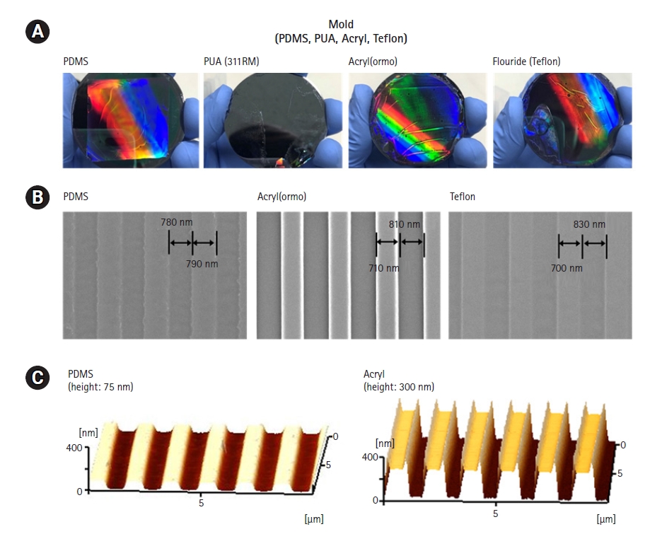
The films were also observed by optical microscopy to visualize large areas (Fig. 2A). In this process, Nafion films were not removed from the base substrate when using a PUA mold. The Nafion films still remained on the base substrate. We suggest that the adhesion force between Nafion and the second substrate was insufficient to remove the Nafion from the base (first) substrate. Some destruction was also observed on the Nafion films when using the Teflon mold. The PDMS and acryl molds showed the best performance on optical microscopy. SEM and AFM images were observed to optimize the mold (Fig. 2B and 2C). Acryl showed a clear and accurate line pattern on the SEM images. PDMS and Teflon showed an accurate linewidth and space, but the height was different from that of the acryl mold. AFM showed the height differences between the PDMS and acryl molds; the acryl mold had an accurate height, whereas the PDMS mold had a height of 150 nm.
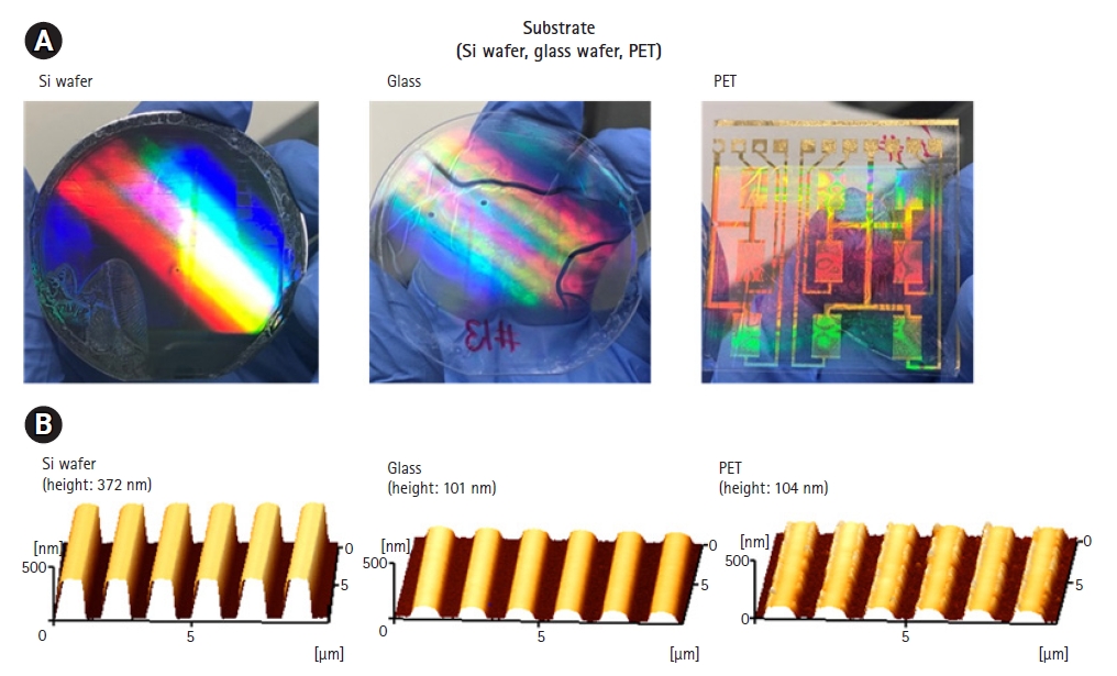
Optical and AFM images of the films on the different substrates are shown in Fig. 3. An advantage of using a Nafion solution, not a Nafion membrane, is that Nafion could be deposited on various substrates. Nafion was deposited on the silicon wafer, the glass wafer, and the PET flexible film using the acryl mold. Due to the optical transparency of the entire structure, Nafion films were applied in optics for fabrication. The lines had a high structural fidelity and resolution, demonstrating the accuracy of this fabrication process to form micro-patterns over large areas [6]. AFM images are shown in Fig. 3B, with patterns showing a linewidth of around 800 nm and a space of 800 nm. However, the silicon wafer had a height of about 400 nm, while the glass wafer and PET film had a height of 100 nm. Thus, the silicon wafer showed the best performance as a substrate to make micro-patterned Nafion films.

2. Evaluation of cell adhesion

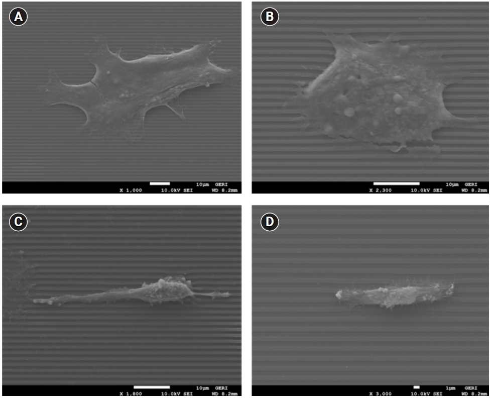
As previously mentioned, the Nafion micro-patterns obtained by the molding process were able to control the cell-adhesive regions, unlike previously reported cell patterning techniques using stamping, enzymes, and other proteins. Fig. 4 shows the cells on films coated with MCF-7 cells (Fig. 4A and 4B) and MDA-MB-231 cells (Fig. 4C and 4D). The cells clearly showed cell adhesion patterns aligning with the designed organization. Most interestingly, although the morphology of cell adhesion of MDF-7 and MDA-MB-231 was different, both MDF-7 and MDA-MB-231 cells migrated from the micro-patterns. These data suggest that the Nafion coating with a micro-pattern organization played a crucial role through its ability to guide and enhance cell growth and adhesion according to a specifically designed order. The cells were guided by the surface morphology and the stiffness differences between the pattern and the substrate. These Nafion films can therefore be used as cell culture sheets for cell-based platforms, where the micro-pattern can be used to guide and align the cells.

Discussion

In this paper, we demonstrate the fabrication of Nafion micro-patterns by molding technology, which permits high resolution, high throughput, and scale. We optimized the micro-pattern using different substrates and mold materials and determined that the acryl mold and silicon substrate showed the best performance. The films are mechanically robust, can be formed at various thicknesses, ranging from 100 nm to 500 nm, and have controllable thickness and pattern spaces. Next, cell adhesion was evaluated using both MCF-7 and MDA-MB-231 cells. These micro-patterned Nafion films not only enhanced cell adhesion, but also facilitated cell migration and alignment. These results suggest that micro-patterned Nafion sheets can serve as a valuable tool for flexible cell-based platforms and devices.

NOTES

Conflict of interest

No potential conflict of interest relevant to this article was reported.

Funding

This work was supported by an Institute of Information & Communications Technology Planning & Evaluation (IITP) grant funded by the Korean government (MSIT) (No. 2021-0-01302, Rollto-plate-based large-scaled drug screening system development, 50%) and National R&D Nano•Material Technology Development Program through the National Research Foundation of Korea (NRF) funded by the Ministry of Science and ICT (No. NRF-2021M3H4A4079264, NRF-2021M3H4A4079275, 30%) and platform engineering by the Small and Medium Business Administration (No. S3310771, 20%).

Authors’ contributions

Conceptualization: SHL; Data curation: HBP, SKS, HJK; Methodology: SHL, HBP, SKS; Supervision: JKL, HJK; Writing-original draft: JKL; Writing-review & editing: all authors.

Data availability

Please contact the corresponding author for data availability.

Fig. 1.
(A) Design and fabrication of the micro-patterned Nafion film. A polyurethane-acrylate (PUA) mold was used to generate a micro-pattern on the Nafion substrate. (B) Scanning electron microscopy images of the micro-patterns on different molds. Polydimethylsiloxane (PDMS), PUA, acryl, and Teflon were used as mold materials, respectively. PET, polyethylene terephthalate.
organoid-2023-3-e8f1.jpg
Fig. 2.
(A) Optical morphology of micro-patterned Nafion films on different molds (PDMS, PUA, acryl, and Teflon). (B) SEM images of micro-patterns on PDMS, acryl, and Teflon. (C) AFM images of the micro-patterned Nafion films. PDMS, polydimethylsiloxane; PUA, polyurethane-acrylate; SEM, scanning electron microscopy; AFM, atomic force microscopy.
organoid-2023-3-e8f2.jpg
Fig. 3.
(A) Optical morphology of the micro-patterned Nafion films on different substrates (Si wafer, glass wafer, and PET). (B) AFM images of the micro-pattern Nafion films on different substrates. An acryl mold was used to fabricate the Nafion film. AFM, atomic force microscopy; PET, polyethylene terephthalate.
organoid-2023-3-e8f3.jpg
Fig. 4.
Scanning electron microscopy (SEM) images of cell adhesion to micro-patterned Nafion films. Cell adhesion in a humidified 5% CO2 atmosphere at 37°C was monitored for 4 days. (A, B) MCF-7, (C, D) MDA-MB 231.
organoid-2023-3-e8f4.jpg

REFERENCES

1. Dobkowski J, Kolos R, Kamiński J, Kowalczyńska HM. Cell adhesion to polymeric surfaces: experimental study and simple theoretical approach. J Biomed Mater Res 1999;47:234-42.
crossref pmid
2. Horbett TA. The role of adsorbed proteins in animal cell adhesion. Colloids Surf B Biointerfaces 1994;2:225-40.
crossref
3. Boyan BD, Hummert TW, Dean DD, Schwartz Z. Role of material surfaces in regulating bone and cartilage cell response. Biomaterials 1996;17:137-46.
crossref pmid
4. Liu VA, Jastromb WE, Bhatia SN. Engineering protein and cell adhesivity using PEO-terminated triblock polymers. J Biomed Mater Res 2002;60:126-34.
crossref pmid
5. Evans MD, Steele JG. Polymer surface chemistry and a novel attachment mechanism in corneal epithelial cells. J Biomed Mater Res 1998;40:621-30.
crossref pmid
6. Xu M, Pradhan S, Agostinacchio F, Pal RK, Greco G, Mazzolai B, et al. Easy, scalable, robust, micropatterned silk fibroin cell substrates. Adv Mater Interfaces 2019;6:1801822.
crossref pdf
7. Kim DH, Lu N, Ma R, Kim YS, Kim RH, Wang S, et al. Epidermal electronics. Science 2011;333:838-43.
crossref pmid
8. Sugano J, Fujie T, Iwata H, Iwase E. Measurement of conformability and adhesion energy of polymeric ultrathin film to skin model. Jpn J Appl Phys 2018;57:06HJ04.
crossref pdf
9. Feinberg AW, Feigel A, Shevkoplyas SS, Sheehy S, Whitesides GM, Parker KK. Muscular thin films for building actuators and powering devices. Science 2007;317:1366-70.
crossref pmid
10. Ogaki R, Alexander M, Kingshott P. Chemical patterning in biointerface science. Mater Today 2010;13:22-35.
crossref
11. Cavalcanti-Adam EA, Volberg T, Micoulet A, Kessler H, Geiger B, Spatz JP. Cell spreading and focal adhesion dynamics are regulated by spacing of integrin ligands. Biophys J 2007;92:2964-74.
crossref pmid pmc
12. Huang J, Grater SV, Corbellini F, Rinck S, Bock E, Kemkemer R, et al. Impact of order and disorder in RGD nanopatterns on cell adhesion. Nano Lett 2009;9:1111-6.
crossref pmid pmc
13. Teixeira AI, Abrams GA, Bertics PJ, Murphy CJ, Nealey PF. Epithelial contact guidance on well-defined micro- and nanostructured substrates. J Cell Sci 2003;116(Pt 10):1881-92.
crossref pmid pmc pdf
14. Srituravanich W, Fang N, Sun C, Luo Q, Zhang X. Plasmonic nanolithography. Nano Lett 2004;4:1085-8.
crossref
15. Manso M, Rossini P, Malerba I, Valsesia A, Gribaldo L, Ceccone G, et al. Combination of ion beam stabilisation, plasma etching and plasma deposition for the development of tissue engineering micropatterned supports. J Biomater Sci Polym Ed 2004;15:161-72.
crossref pmid
16. Delamarche E, Bernard A, Schmid H, Bietsch A, Michel B, Biebuyck H. Microfluidic networks for chemical patterning of substrates: design and application to bioassays. J Am Chem Soc 1998;120:500-8.
crossref
17. Javaherian S, O'Donnell KA, McGuigan AP. A fast and accessible methodology for micro-patterning cells on standard culture substrates using Parafilm™ inserts. PLoS One 2011;6:e20909.
crossref pmid pmc
18. Carrico IS, Maskarinec SA, Heilshorn SC, Mock ML, Liu JC, Nowatzki PJ, et al. Lithographic patterning of photoreactive cell-adhesive proteins. J Am Chem Soc 2007;129:4874-5.
crossref pmid pmc
19. Shelly M, Lee SI, Suarato G, Meng Y, Pautot S. Photolithography-based substrate microfabrication for patterning Semaphorin 3A to study neuronal development. Methods Mol Biol 2017;1493:321-43.
crossref pmid pmc
20. Xia Y, Whitesides GM. Soft Lithography. Angew Chem Int Ed Engl 1998;37:550-75.
crossref pmid
21. Roth EA, Xu T, Das M, Gregory C, Hickman JJ, Boland T. Inkjet printing for high-throughput cell patterning. Biomaterials 2004;25:3707-15.
crossref pmid
22. Haraguchi Y, Shimizu T, Yamato M, Okano T. Scaffold-free tissue engineering using cell sheet technology. RSC Adv 2012;2:2184-90.
crossref
23. Ke Q, Wang X, Gao Q, Wu Z, Wan P, Zhan W, et al. Carrier-free epithelial cell sheets prepared by enzymatic degradation of collagen gel. J Tissue Eng Regen Med 2011;5:138-45.
crossref pmid
24. Fujie T, Ahadian S, Liu H, Chang H, Ostrovidov S, Wu H, et al. Engineered nanomembranes for directing cellular organization toward flexible biodevices. Nano Lett 2013;13:3185-92.
crossref pmid
25. Fujie T, Desii A, Ventrelli L, Mazzolai B, Mattoli V. Inkjet printing of protein microarrays on freestanding polymeric nanofilms for spatio-selective cell culture environment. Biomed Microdevices 2012;14:1069-76.
crossref pmid pdf
26. Gelbard G. Organic synthesis by catalysis with ion-exchange resins. Ind Eng Chem Res 2005;44:8468-98.
crossref
27. Cho HD, Won J, Ha HY, Kang YS. Nafion composite membranes containing rod-shaped polyrotaxanes for direct methanol fuel cells. Macromol Res 2006;14:214-9.
crossref pdf
28. Steele JG, Johnson G, Norris WD, Underwood PA. Adhesion and growth of cultured human endothelial cells on perfluorosulphonate: role of vitronectin and fibronectin in cell attachment. Biomaterials 1991;12:531-9.
crossref pmid
29. Fujie T. Development of free-standing polymer nanosheets for advanced medical and health-care applications. Polym J 2016;48:773-80.
crossref pdf
TOOLS
METRICS Graph View
  • 0 Crossref
  •  0 Scopus
  • 6,417 View
  • 33 Download
ORCID iDs

Hyung Jin Kim
https://orcid.org/0000-0001-6766-7625

Related articles


ABOUT
BROWSE ARTICLES
EDITORIAL POLICY
FOR CONTRIBUTORS
Editorial Office
Room 319, Hall 1 of Chonbuk National University Dental College, 20, Geonji-ro, Deokjin-gu, Jeonju 54907, Korea
Tel: +82-63-270-4024    E-mail: editor@j-organoid.org                

Copyright © 2024 by The Organoid Society.

Developed in M2PI

Close layer
prev next