


Introduction
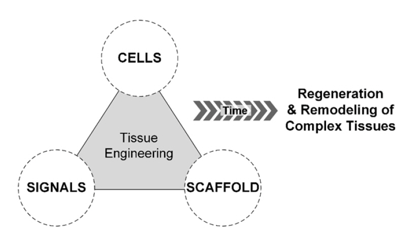
The quest for longevity and improved quality of life has driven mankind to seek definitive solutions to health issues. The first successful organ transplant without immune rejection in 1954 marked a significant milestone in medical history [1]. This revolutionary procedure offered a lifeline to patients burdened with malfunctioning organs. However, the demand for organs has consistently exceeded the available supply, resulting in lengthy waiting lists for many patients [2]. This critical shortage has spurred the development of a new field: tissue engineering. This discipline aims to address the organ shortage by reconstructing, repairing, and regenerating damaged tissues and organs. This is accomplished by using cells as building blocks, scaffolds as structural matrices, and biological cues as essential signals for organ and tissue formation (Fig. 1) [3].
The term “tissue engineering” was first coined by Fung in 1987, marking a significant milestone in the inception of this innovative field of study [4]. Tissue engineering is a multidisciplinary field that combines advanced areas of study such as biology, materials science, chemical engineering, mechanical engineering, and bioengineering. The ultimate objective of tissue engineering is to fabricate tissues and organs for transplantation by replicating living systems, leveraging a profound understanding of the leading technologies from these fields. However, the human body possesses a highly organized and intricately coordinated microenvironment, which is challenging to replicate in vitro. Initially, researchers attempted to emulate the complexity of the human body in a plastic dish by co-culturing a combination of heterotypic cells. Subsequently, researchers began exploring 3-dimensional (3D) culturing methods to bridge the gap between 2-dimensional (2D) cell culture systems and native human body systems. As a result, contemporary advancements in tissue engineering are moving towards the development of 3D constructs with complex microenvironments. This progression is supported by collective accomplishments across various disciplines, including materials science, mechanical engineering, and stem cell technologies, among others [5-7].
Bioprinting is an emerging technology that can be used to create a 3D tissue model of a complex structure [8,9]. This technology facilitates the automated placement of cells and biomaterials in 3 dimensions, enabling the precise and tailored fabrication of tissue models. Bioprinting techniques have been utilized in a variety of tissues to date, including skin, cornea, retina, liver, blood vessels, airways, cartilage, heart, and bladder [10-18]. The use of bioprinting in tissue engineering could lead to more precise models for in vitro drug screening and toxicity research. Among the various bioprinting technologies, piezoelectric inkjet bioprinting is particularly well-suited for replicating the properties of thin and spatially complex soft tissues. This is due to the drop-on-demand (DOD) printing method’s advantages over other bioprinting techniques, such as high resolution, rapid printing speed, high cell viability, and minimal material waste. In this review, we will concentrate on the principles and applications of inkjet-based bioprinting technology.
Technologies for manipulation of cells in 2D
Ethics statement: This study was a literature review of previously published studies and was therefore exempt from institutional review board approval.
The concept of keeping cells alive while being apart from the host organism was established at the end of the 18th century [19]. In 1885, Roux demonstrated that embryonic chick cells could survive in a warm saline solution. Later, in 1916, Rous introduced the use of trypsin treatment for cell suspension. Additionally, in 1921, Carrel reported that cells could be cultivated for extended periods under nutritious and sterile conditions. These breakthroughs in in vitro cell cultivation represent some of the most revolutionary advancements in the field of life science. They have provided an effective tool for monitoring cellular reactions in vitro, a practice that continues to this day.
Cell-to-cell interactions play a crucial role in both developmental stages and adult physiology, making the co-culturing of heterotypic cells a widely used technique [20]. The complexity that arises from co-cultivating cells provides a deeper understanding of cellular characterization and functions. For example, co-cultivating hepatocytes with other cells has been shown to stabilize the viability and function of the hepatocytes more than when they are cultured alone [21]. There are several well-known conventional methods for co-cultivating cells. These typically involve either randomly mixing multiple types of cells at a specific ratio or seeding them separately in a transwell system, which enables compartmentalization of the growth area while still sharing the conditioned media. However, under traditional culture conditions, cells can lose the spatial, chemical, and mechanical information of their in situ cellular microenvironment [22].
Since the late 1900s, researchers have begun to understand the complex in vivo microenvironment through the micropatterning of cells, a significant advancement from traditional randomly distributed co-cultures. They have micropatterned living cells into predetermined designs in vitro to mimic the intricate microenvironment found within our bodies. Notable cell micropatterning methods include photolithography [23-25], soft lithography [26-29], and microfluidics [30-33]. Photolithography techniques employ a series of ultraviolet treatments and toxic solvents to create sub-micrometer-scale patterns on a surface, which then allows cells and proteins to adhere. Soft lithography techniques, on the other hand, utilize bio-friendly soft elastomeric stamps to produce patterns ranging from micrometer to nanometer scale. However, multiple mold fabrication steps are necessary to create these stamps. Microfluidic channels provide 3D dynamic flows to form patterns and facilitate the simultaneous patterning of multiple materials. The complexity of these patterns is directly related to the number of channels, which are created through a complex fabrication process. These traditional micropatterning methods have allowed for more detailed and in-depth investigations into cellular behaviors, biological phenomena, and the interactions between a physiological environment and cells, compared to standard 2D cell cultures. However, these methods involve intricate and time-consuming fabrication steps and necessitate a significant amount of technical knowledge or experience with surface engineering using harmful organic materials. Another challenge lies in accurately positioning multiple cell types on the same substrate using various design patterns.
Bioprinting
Bioprinting is a sophisticated tool that allows for the automated and computer-assisted spatial placement of cells. This technology employs a bottom-up approach, eliminating the need for external scaffolding during the fabrication process. Its ability to deposit cells and biomaterials according to a predetermined design makes it highly efficient for creating 2D cell micropatterns and 3D cell-embedded constructs. Two-dimensional cell micropatterns can be fabricated by bioprinting cell adhesive proteins or by directly bioprinting the cells themselves [34-36]. Table 1 presents a comparison between conventional methods and bioprinting for the fabrication of 2D cell micropatterns. Unlike traditional scaffolding methods, bioprinting allows for the simultaneous positioning of cells during the fabrication of 3D cell-laden constructs.
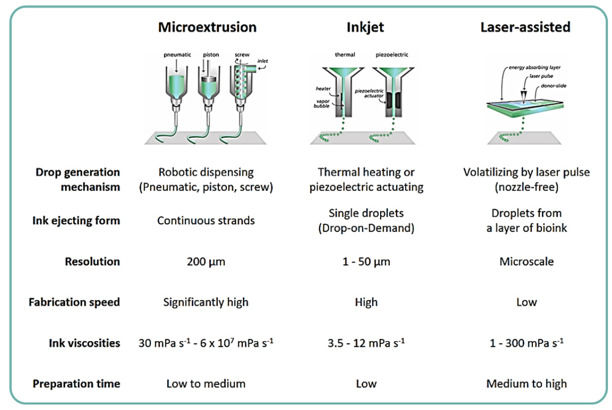
The primary subtypes of bioprinting technology include microextrusion [37-40], inkjets [35,41-48], and lasers (Fig. 2) [49-53]. Microextrusion bioprinting is the most commonly used and cost-effective technology. This system dispenses bioink through a nozzle using either pneumatic or mechanical piston- and screw-type operational modules. While microextrusion bioprinters offer high-speed and straightforward deposition, their low resolution is a notable limitation [9,54]. Inkjet bioprinting encompasses both thermal bubble jet and piezoelectric types. This system deposits bioink through a nozzle in small picoliter-volume droplets using a DOD method. The drop-generating mechanism of inkjet printing allows for high resolution, close to 50 μm [55]. High-speed printing is also possible, but the inkjet printers can only use inks with liquid-like and less-viscous properties [56]. Laser-assisted bioprinting employs laser energy to vaporize and propel a sacrificial layer, producing droplets onto a receiving substrate. This system achieves high resolution at a microscale using a nozzle-free approach. However, this high-resolution performance results in a relatively low overall flow rate, making the process time-consuming and hindering the fabrication of applicable 3D constructs [53]. Each of these bioprinting types has distinct features and advantages. Therefore, careful consideration should be given to the selection of an appropriate bioprinting method or the hybridization of multiple bioprinting methods.
Principles of inkjet printing
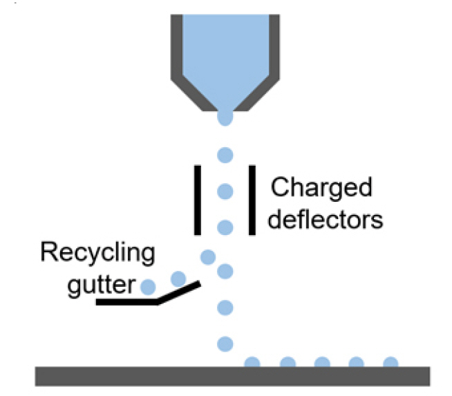
Digital inkjet printing is an extensively researched additive printing process that has gained popularity due to its unique DOD approach. This method eliminates waste from materials and premanufactured master printing plates [57-61]. Continuous inkjet printing, on the other hand, generates ink drops in a continuous stream at an operational frequency of 20 to 60 kHz, even when printing is not necessary (Fig. 3). To minimize ink waste, unwanted drops can be deflected into a gutter by electrically charged deflectors and then recycled. As a result, continuous inkjet printing is widely employed in the industrial sector for efficient product marking. In contrast, DOD type inkjet printers can eject single drops at the picoliter level only when needed. With these compelling advantages, inkjet printing technology has long been a powerful tool in the field of printed electronics, such as organic thin-film transistors and light-emitting devices [62]. Additionally, inkjet printing technology has found applications in the biomedical field, including biosensors and micro-arrays of DNA or proteins [63].
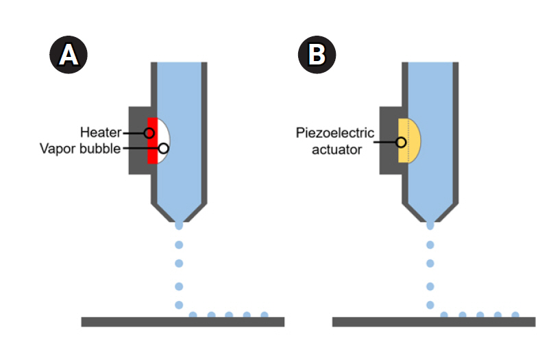
The print heads of DOD inkjet printers are primarily categorized into 2 distinct groups: the thermal bubble jet type and the piezoelectric type (Fig. 4) [64]. The thin-film heater within the ink chamber of the bubble jet is heated to generate bubbles, which then eject fluidic drops from the nozzle’s orifice. Conversely, the piezoelectric type of inkjet head contains a piezoelectric transducer within the ink chamber, which transforms electrical signal inputs into mechanical deformation. Drops are printed from the ink chamber due to the volumetric change resulting from the mechanical actuation of the piezoelectric material. As such, the generation of drops, including their size, velocity, and morphology, can be modulated by designing actuation pulses. The size of the drops primarily depends on the diameter of the orifice, but it is also influenced by the applied pulse. The typical size of inkjet printed drops ranges between 10 to 150 μm, with a volume at the picoliter level. Drops can be ejected at frequencies ranging from 1 to 20 kHz.
Inkjet-based 2D cell patterning
Living cells have been micropatterned into predetermined designs in a laboratory setting to replicate the intricate microenvironment found within our bodies. Representative methods of cell micropatterning include photolithography, soft lithography, and microfluidics. These traditional micropatterning techniques have facilitated more detailed and in-depth studies of cellular behaviors, biological phenomena, and the interactions between a physiological environment and cells, compared to standard 2D cell culture. However, these methods entail complex and lengthy fabrication processes and necessitate significant technical knowledge or experience in surface engineering using potentially harmful organic materials. An additional challenge lies in accurately positioning multiple cell types on the same substrate using a variety of design patterns.
Bioprinting has recently emerged as a promising micropatterning technology, utilizing a bottom-up and scaffold-free fabrication approach. The first instance of inkjet bioprinting was reported by Wilson and Boland in 2003 [65], who used living cells and biomaterials as ink in a commercial inkjet printer. Modifications were made to the print heads, printer hardware, and software of the commercial HP inkjet printer to enable the delivery of protein or cell solutions for a variety of applications. This innovative approach paved the way for a new strategy in fabricating cell patterns, distinct from traditional photolithography or soft lithography methods. The majority of initial studies on inkjet bioprinting focused on printing cell-adhesive or cell-repellent materials onto a substrate, followed by manual seeding of living cells to create cell patterns [35,41,45]. Cell-repellent materials such as poly(ethylene) glycol (PEG), alginate, and agarose were commonly used to coat bare glass backgrounds before subsequent inkjet printing of cell-adherent materials. Materials typically used for cell adhesion include collagen, gelatin, hyaluronic acid, poly-D-lysine, poly-L-lysine, and poly(lactic-co-glycolic acid). The inkjet printing of cell adhesive or repellent ink compartmentalized the surface for cells, resulting in the fabrication of precise 2D cell patterns
Meanwhile, living cells have been directly inkjet-printed onto substrates. In 2005, mammalian cells, including Chinese hamster ovary and embryonic motoneuron cells, were directly printed using a modified HP thermal inkjet printer, achieving a viability rate of over 90% [46]. An electrostatically driven inkjet system, which did not generate heat in the ink chamber, was also employed to deposit patterns of bovine endothelial cells [66]. Human fibroblasts, HT 1080, were printed using a piezoelectric inkjet printer, demonstrating a survival rate comparable to that of thermal inkjet-printed cells [67]. Furthermore, studies have been conducted to optimize printing conditions for single-cell level manipulation. For this type of manipulation using inkjet printing, images of the nozzle orifice were analyzed before ejection [68,69], an additional oil layer was applied to segregate printed cell suspensions into individual wells [70], probe electrospray ionization mass spectrometry was adapted to capture cells under an electrical field [71], and a nozzle with a small diameter (30-μm), similar to the size of cells, was chosen [72]. Inkjet printing for cell-level manipulation can be applied to biomedical applications such as high-throughput screening and biofabrication with controlled cell numbers.
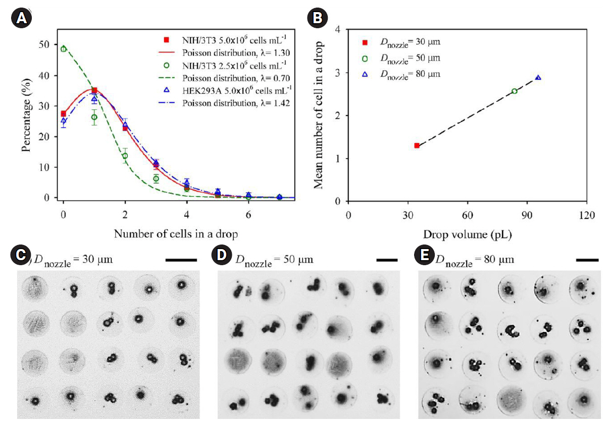
More recently, the precision of mammalian cell printing has been significantly improved through the use of a DOD inkjet with a 30-μm nozzle diameter, allowing for cell-level accuracy [72]. High-speed imaging methods were employed to investigate the motion of cells within the nozzle under pulsed pressure generated by a piezoelectric transducer, as well as the formation of jets after cell ejection. Fig. 5 demonstrates the influence of cell type, cell concentration, and nozzle diameter on the number of cells contained in a single droplet [72]. This was exemplified by printing patterns of 20×20 dot arrays on a glass substrate. To further illustrate this relationship, representative microscopic images of printed droplets from various nozzle sizes were provided. The study found that the 30-μm inkjet nozzle had minimal impact on the printed mammalian cells, causing negligible damage.
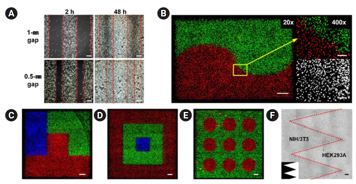
For the first time, Park et al. [73] introduced a DOD inkjet printing method that directly patterned living cells into a cell-friendly liquid environment [74]. They meticulously fine-tuned the printing parameters and used high-speed imaging to observe the behaviors of cell jetting and impacting, achieving precise control over cell placement with exceptional resolution. This allowed them to showcase the capabilities of the direct cell printing method, as they successfully co-printed various types of cells into a range of designs, including complex gradient arrangements (Fig. 6) [73]. Lastly, they applied the cell printing method to study the impact of heterogeneity and geometry of the cell population on the infectivity of the seasonal H1N1 influenza virus (PR8). This demonstrated that direct inkjet cell patterning can serve as a powerful and versatile tool for both fundamental biology and applied biotechnology.
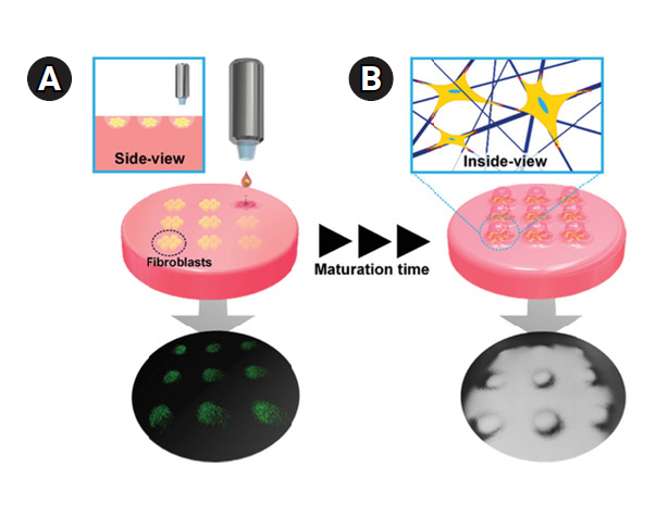
The team further refined the inkjet-based cell patterning technique, devising a strategy for the self-organization of 3D collagen microstructures [75]. Fig. 7 illustrates the use of a DOD inkjet printer to generate patterned fibroblast cells on a collagen substrate, adhering to predetermined patterns and regulated density [75]. The study’s results suggest that the interaction between cells and the extracellular matrix (ECM) promotes the self-organization of cellular structures within the collagen hydrogel, as depicted in the accompanying figure. By leveraging this mechanism, the dimensions and shapes of the 3D collagen microstructures were regulated by adjusting the cell pattern designs and cell density. Ultimately, this method was employed to construct a human skin model featuring papillary microstructures at the dermo-epidermal junction.
Hydrogels for inkjet bioprinting
Various hydrogels with hydrophilic polymer chains, such as alginate, collagen and gelatin methacrylate, are widely used in inkjet bioprinting. Alginate is frequently employed to construct scaffolds with calcium chloride (CaCl2) as a cross-linker. Xu et al. [76] proposed a platform-assisted 3D inkjet bioprinting system to develop complex 3D constructs such as zigzag tubes. In this system, the XY stages were meticulously controlled to set the dispensing head's location for planar feature printing. Simultaneously, the Z stage was systematically adjusted to match the printing speed for each layer (Fig. 8) [76]. After printing a layer, the platform attached to the Z stage must descend by a distance equal to the layer’s thickness to allow gelation of each newly deposited layer. Xu et al. [43] created pie-shaped multicellular heterogeneous tissue constructs by directly and simultaneously inkjet printing multiple cell types. They separately mixed human amniotic fluid-derived stem cells, canine smooth muscle cells, and bovine aortic endothelial cells with the ionic cross-linker CaCl2 and printed them using a thermal inkjet printer. During the bioprinting process, the 3 different cell types were strategically placed in specific positions within a chamber filled with a sodium alginate-collagen composite material. The resulting 3D constructs, produced using this bioprinting technique, demonstrated the potential to flourish and mature into fully functional tissues with adequate vascularization in vivo.
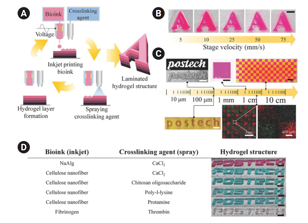
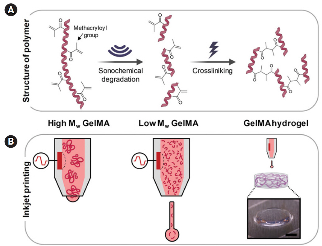
Yoon et al. [77] developed a novel bioprinting method that combines DOD inkjet printing with a spray-coating technique. This approach allows for the high-resolution, high-speed, and freeform creation of large-scale structures made of cell-laden hydrogel (Fig. 9). They employed this hybrid inkjet-spray printing technique to construct 3D biological hydrogel structures in various shapes, using materials such as alginate, cellulose nanofiber, and fibrinogen. Their research demonstrated that the inkjet-spray printing system effectively facilitated the production of cell-laden hydrogel structures with remarkable shape accuracy, while also providing a quick and dependable manufacturing process. More recently, they introduced the sonochemical degradation of gelatin methacryloyl (GelMA) as a means to regulate the viscoelasticity of a GelMA-based bioink. This is achieved by reducing the length of the polymer chains without chemically damaging the methacryloyl groups (Fig. 10) [78]. By adopting this approach, the maximum attainable polymer concentration for printing is significantly increased, rising from 3% to 10%. This heightened control over the mechanical properties of GelMA hydrogels could potentially encourage better fibroblast spreading on the hydrogels. Additionally, they successfully created 3D constructs composed of multiple layers of cell-laden hydrogels with distinct physical properties, using high-resolution inkjet printing technology.
Inkjet-printed tissues and organs
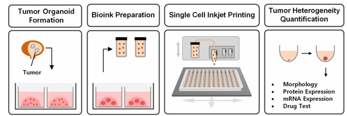
In this section, we review representative examples of inkjet-printed tissues and organs for future regenerative medicine, toxicology, and pharmaceuticals. As one example, a piezoelectric inkjet printer was used to analyze intratumoral heterogeneity in bladder cancer for the first time [79]. Cells dissociated from patient-derived tumor organoids were inkjet-printed, allowing for precise distribution into a microwell plate without the need for additives or treatments. These cells were then cultured into organoids for further analysis, as depicted in Fig. 11 [79]. The mRNA expression levels of representative luminal and basal genes in both types of tumor organoids were assessed to confirm the heterogeneous expression of different genes among individual organoids. The quantification of intratumoral heterogeneity is crucial for the development of effective therapeutic strategies in the age of personalized medicine. This study illustrates that the fully automated inkjet printing system can serve as a valuable tool for cell sorting and quantifying intratumoral heterogeneity.
Inkjet bioprinting is a promising technology for fabricating skin substitutes and has the potential to revolutionize wound healing and skin grafting procedures. Lee et al. [80] utilized a pneumatic microextrusion technique to create a sturdy dermal layer composed of collagen and primary human dermal fibroblasts. Following the creation of this dermal layer, they employed piezoelectric inkjet printing to deposit a layer of primary human epidermal keratinocytes. Their goal was to establish an epidermal layer that exhibited both high cellular viability and uniformity (Fig. 12). They then used these 3D bioprinted skin models to examine the effects of Aronia melanocarpa extract on human skin condition. Their research, facilitated by the development of 3D skin models, revealed that the Aronia extract enhanced the synthesis of type I collagen and reduced the expression of MMP1 and MMP3. These findings suggest that this extract could be beneficial in treating damaged or aged skin [81].
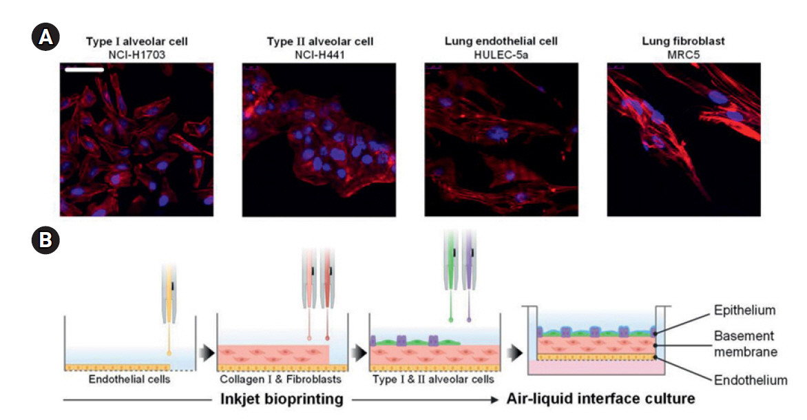
Physiologically relevant models of the human respiratory system are urgently needed to study disease pathogenesis and drug efficacy, particularly in light of the emergence of new respiratory viruses and the high mortality rates associated with lung diseases. Kang et al. [82] has reported the use of automated high-resolution inkjet printing to deposit alveolar cells, which has allowed for the creation of a 3-layered alveolar barrier model with an unprecedented thickness of approximately 10 μm (Fig. 13) [82]. Their research demonstrated that this 3D structured model more accurately replicated the structure, morphology, and functions of lung tissue. This was not only in comparison to a traditional 2D cell culture model, as one might expect, but also when compared to an unstructured 3D model consisting of a homogeneous mixture of alveolar cells and collagen. Lastly, it was found that this thin, multilayered model effectively mimics the tissue-level responses to influenza infection.
This model was employed to investigate the impact of fine dust particles on respiratory diseases. The 3-layered in vitro model was subjected to A2 fine test dust at varying concentrations and for different durations [83]. The effects observed in the alveolar tissue exposed to high concentrations of fine dust particles included: (1) an elevated expression of pro-inflammatory cytokines, (2) damage and disarray of tissue structure, and (3) the up- or downregulation of genes linked to respiratory diseases. The study demonstrated that a 3D-printed tissue model can be used to assess the physiological effects of fine dust particles on cytotoxicity, the rigidity of the alveolar barrier, and the secretion of surfactant from the alveolar barrier. Additionally, a pulmonary fibrosis model was created by treating the 3D alveolar barrier with a pro-fibrotic cytokine. The suitability of this model for studying fibrosis was confirmed by observing changes in structural deposition, pulmonary function, epithelial-mesenchymal transition, and fibrosis markers [83].
The lung-on-a-chip is an innovative platform for cell culture, designed to replicate the physiological microenvironment of the lungs, including perfusion and lung functionality [84-86]. Huh et al. [87] developed a representative lung-on-a-chip model that mimics the mechanical and structural properties of the alveolar barrier. This model uses a microfluidic device with a porous polymer membrane to create the alveolar-capillary interface by seeding epithelial and endothelial cells on opposite sides. There have been significant efforts to improve this approach and create complex tissues that mirror the physiological composition of the lungs. For example, researchers have segmented fibroblasts and smooth muscle cells, arranging them in multilayered microfluidic compartments to replicate the intricate nature of lung tissue [88,89]. In other studies, the synthetic porous membrane has been replaced with a matrix-driven membrane [90-93] or a native ECM structure [94]. Recent research has concentrated on developing advanced membranes that closely mimic the mechanical characteristics of the lungs, such as thickness and flexibility [95-97]. However, most tissue fabrication methods involve infusing a high-density cell suspension into a microfluidic channel, where cell sedimentation determines their positioning and overall tissue structure. This indirect cell seeding process has limitations in accurately replicating the ultra-thin and multilayered barrier architecture, as well as achieving precise spatial arrangements of multiple cell types within the unique structure. Additionally, ensuring high repeatability and uniform thickness in tissue fabrication remains a challenge.
To address these limitations, Kim et al. [98] developed a human alveolar lung-on-a-chip model that closely replicates physiological conditions. This model incorporates a micron-thick, 3-layered tissue that is printed using an inkjet printing technique (Fig. 14). The team printed lung tissues sequentially within 4 culture inserts, then implanted these into a biochip that ensures a steady flow of culture medium. This modular implantation process led to the creation of a lung-on-a-chip system, which enables the growth of inkjet-bioprinted lung models in a 3D structure, while maintaining perfusion at the air-liquid interface. The bioprinted models, cultured on the chip, retain their structural integrity. They consist of 3 layers with micrometer-scale thickness and exhibit the formation of tight junctions in the epithelial layer.
Summary and outlook
In summary, inkjet bioprinting has emerged as a significant tool in the fields of tissue engineering and regenerative medicine. This technique's capacity to produce picoliter volume droplets swiftly, without contact, and with high precision, has rendered it an invaluable resource in the creation of complex two- 2D and 3D biological constructs. The evolution of this technology is evident in its applications, shifting from its traditional use in the publishing industry to its current role in modern medicine for drug screening, disease modeling, and toxicology testing, as previously discussed. As this technique continues to develop, we expect a substantial increase in the precision and complexity of bioprinted tissues and organs. With further advancements in smart biomaterials and fabrication methods, inkjet bioprinting could enable the production of dynamic 3D structures that can respond to biological cues, thus more accurately replicating native biological systems.


 PDF Links
 PubReader
 ePub Link
 Full text via DOI
 Download Citation
 Print
Print


