Organoid > Volume 4; 2024 > Article
Yang, Kiral, and Park: Telencephalic organoids as model systems to study cortical development and diseases

Abstract

The telencephalon is the largest region of the brain and processes critical brain activity. Despite much progress, our understanding of the telencephalon’s function, development, and pathophysiological processes remains largely incomplete. Recently, 3-dimensional brain models, known as brain organoids, have attracted considerable attention in modern neurobiological research. Brain organoids have been proven to be valuable for studying the neurodevelopmental principles and pathophysiology of the brain, as well as for developing potential therapeutics. Brain organoids can change the paradigm of current research, replacing animal models. However, there are still limitations, and efforts are needed to improve brain organoid models. In this review, we provide an overview of the development and function of the telencephalon, as well as the techniques and scientific methods used to create fully developed telencephalon organoids. Additionally, we explore the limitations and challenges of current brain organoids and potential future advancements.

Introduction

The telencephalon comprises major areas in the mammalian brain with several important components, including the cerebral cortex, limbic system, basal ganglia, and the olfactory system [1,2]. The development of the telencephalon, which has several distinct parts, requires an interplay of diverse signaling pathways that are tightly regulated from the embryonic to the adult stages. In addition, various diseases related to telencephalon development arise due to genetic mutations or external factors [3]. Despite significant progress over the past decades in uncovering the mechanisms of brain development and pathophysiology, the intricate structure and function of the brain present a major challenge. Recently, models known as brain organoids have been developed to mimic the developing human brain [4]. Brain organoid technologies are excellent platforms for probing brain development, pathophysiology, and mechanisms. In this review paper, we will provide a brief overview of telencephalon development and telencephalon brain organoids, and we will also discuss the limitations of the current organoid system and future perspectives.

The telencephalon: an overview

Ethics statement: This study constituted a comprehensive analysis of previously released studies and thus, was not subject to the approval of the institutional review board.
The forebrain, also called the prosencephalon, comprises the largest part of the brain. It plays a key role in sensory processing, perception, and cognitive functions related to information processing [5,6]. The forebrain is divided into 2 regions: the telencephalon and the diencephalon. The telencephalon occupies the largest part of the central nervous system [7]. It is responsible for olfactory processing as well as speech, language, and memory formation. The main component of the telencephalon is the cerebral cortex, which is further divided into 4 lobes: the frontal, parietal, occipital, and temporal lobes [6]. The diencephalon is divided into 3 parts (the thalamus, epithalamus, and subthalamus) and has the function of maintaining homeostasis in the body [7]. The hypothalamus arises developmentally from the telencephalon, while anatomically it is adjacent to the regions from the diencephalon. Overall, the telencephalon and diencephalon have different roles, but they closely interact to perform their essential functions.
The frontal lobe, which is located at the front of the cerebrum, governs higher mental functions such as memory, thinking, and reasoning. It processes information from other association areas and regulates behavior. Damage to the frontal lobe often leads to a loss of problem-solving abilities and the capacity to plan and execute actions, such as crossing a street or answering complex questions [8,9]. Frontal lobe syndrome is a common condition associated with this area, and there are instances where a person’s behavior or personality changes due to trauma or various diseases [10]. The temporal lobe is the lateral part of the cortex. The right temporal lobe controls the left side of the body, and the left temporal lobe controls the right side. Functionally, it is mainly responsible for auditory stimulation, language, and emotional response. The medial temporal lobe includes the amygdala and hippocampus, which are the main structures forming the limbic system and play a critical role in memory function [11]. Temporal lobe epilepsy is one of the most common types of epilepsy in adults, and it is most frequently caused by sclerosis of the medial temporal lobe, particularly the hippocampus [12]. The occipital lobe is found at the back of the cerebral cortex and is the smallest lobe. Its functions primarily involve processing visual information coming from the eye. The primary visual cortex is the visual center of the occipital lobe. Visual information processed here is divided into 2 pathways: one towards the parietal lobe and the other towards the temporal lobe. The dorsal pathway to the parietal lobe processes visual information about moving objects, such as position, speed, and distance, as well as information about eye and body movements. The ventral pathway to the temporal lobe is responsible for judging the color and shape of the object being viewed by comparing it with existing images, and contributes to the long-term storage of visual memory [13]. Balint’s syndrome, characterized by severe spatial deficits and neuropsychological disorders, is a representative disease associated with this lobe [14]. The parietal lobe, which is located just behind the central sulcus in the cerebral cortex, is responsible for perceiving tactile and spatial senses and responding to the movement of objects in sight. It also integrates information from the outside world, combining letters into words to give them meaning [15,16]. Gerstmann syndrome, which causes learning disabilities and cognitive impairment, is a condition that can occur when the parietal lobe is damaged [17].

Early telencephalon development: a tale of signaling in neurogenesis.

The human brain development process consists of the generation, migration, and differentiation of neurons, as well as the maturation and formation of synapses [18]. Inhibitors of bone morphogenic protein (BMP), secreted from the organizer, induce the ectoderm to transform into nervous tissue, thereby forming a neural plate through morphogenesis [19]. The neural plate then folds in on itself to form the neural tube, with dorsal/ventral and anterior/posterior fates being patterned by the collective influence of signaling molecules (Fig. 1) [20].
In the early stage of embryonic development, the central nervous system is subdivided into the forebrain, midbrain, hindbrain, and spinal cord along the anterior-posterior axis [21]. Various signaling pathways mediate this anterior-posterior patterning. The telencephalon originates from cells at the rostral part of the neural plate [22]. Wnts are known to play an important role in rostral-caudalization; thus, proper regulation of the interaction of Wnts and their antagonists is crucial in the establishment of the telencephalon [23]. Once the anterior and posterior of the neural plate are formed, the telencephalon undergoes dorsal-ventral patterning [24]. The telencephalon in mammals develops as the pallium of the dorsal region and the subpallium of the ventral region by dorsal-ventral patterning. The pallium is further divided into 4 regions: dorsal, medial, lateral, and ventral [25]. The dorsal pallium develops into the neocortex, which has the most complex structure in mammals. The medial pallium gives rise to the medial entorhinal cortex, hippocampus, cortical hem, and choroid plexus. The insular cortex and the lateral entorhinal cortex are known to originate from the lateral pallium. The ventral portion at the pallial-subpallial boundary gives rise to the amygdala [26-28]. In contrast, lateral, medial, and caudal ganglionic eminences (LGE, MGE, and CGE) in the subpallium develop as the basal ganglia [29]. Specific domains of the early telencephalon produce distinct sets of neurons and eventually generate the neural network of the mature telencephalon.
The regional patterning of the telencephalon is regulated by various morphogens, including Wnt, BMP, retinoic acid (RA), sonic hedgehog (Shh), and fibroblast growth factor (FGF). BMP or Wnt signaling is required for dorsal patterning of the telencephalon, while Shh signaling is important for ventral patterning, and RA is crucial for lateral patterning [22,25,30]. Wnts are expressed at the dorsal midline of the telencephalon, known as the cortical hem. Wnt signaling is important for patterning the medial pallium, which later develops into the hippocampus and medial entorhinal cortex [26,31]. When Wnt3a is knocked out and Wnt signaling is lost, the mouse hippocampus does not develop normally [32,33]. Furthermore, the hippocampus develops poorly in LEF1-knockout mice, a transcription factor known as a target gene for Wnt/β-catenin signaling [34]. BMPs are secreted from the lateral edges and dorsal midline of the neural plate. They enhance dorsomedial identity and the development of the choroid plexus [35,36]. The Shh signaling pathway is mediated by Smoothened, which specifies the ventral telencephalon [37]. The major source of Shh is the cells of the floor plate in the neural tube [38]. Under tight control of the gradient of Shh signaling, the ventral telencephalon gives rise to various ganglionic eminences (medial, caudal, and lateral) [39,40]. The ventral telencephalon in Shh-knockout mice displays the absence of expression of the telencephalon ventral markers Nkx2.1, Dlx2, and Gsx2 in neural progenitor cells [41].

Brain organoids: in vitro models of the human brain telencephalon

Over the years, researchers have actively used neural stem cells and neurons in cell culture systems, typically in 2-dimensional (2D) culture formats, or animal models to study the neural system. The use of cells in 2D culture is straightforward for addressing questions directly related to intrinsic cell features. However, this approach does not account for the interaction between the cells and the extracellular substrate found in tissue. Furthermore, the cerebral cortex is primarily composed of 6 laminar layers, a complexity that 2D culture cannot replicate [42]. Animal models have limitations in reproducing human responses due to species differences, necessitating new model systems.
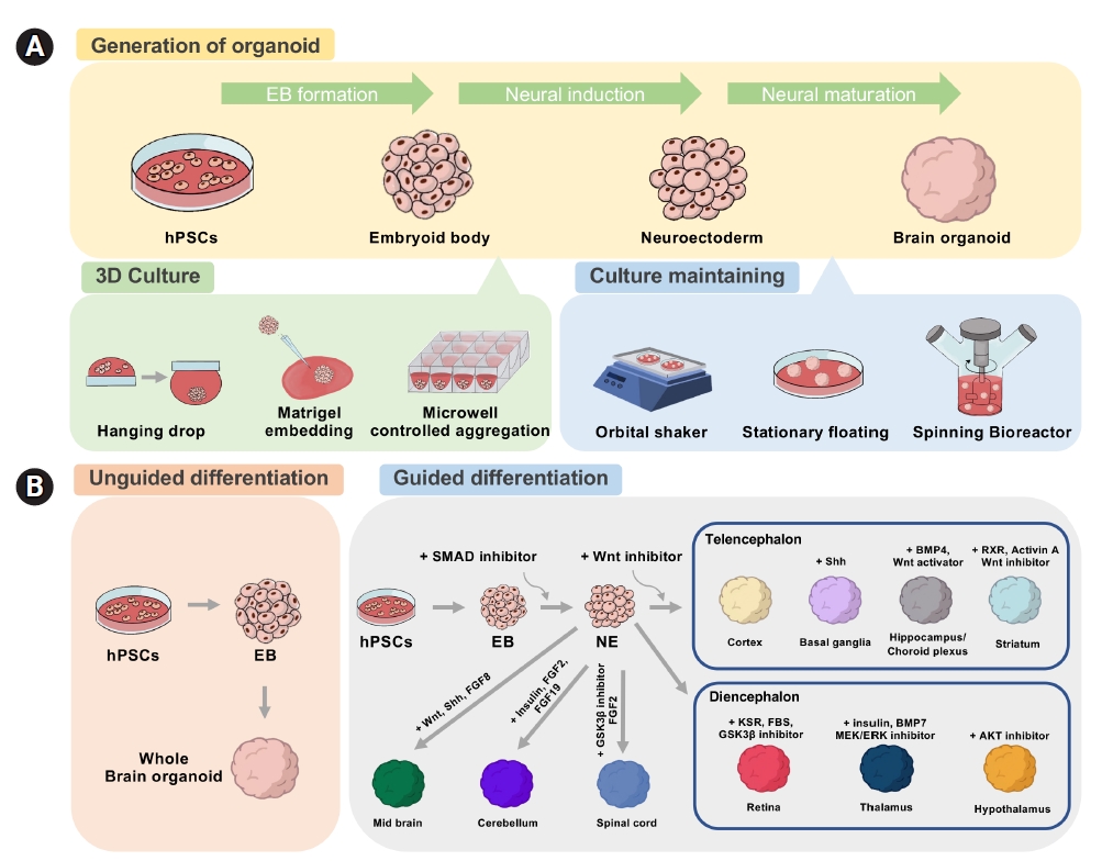
The establishment of stem cell culture conditions and 3-dimensional (3D) culture techniques enabled the development of brain organoids (Fig. 2) [43]. Organoids are constructed by culturing self-organizing cells with multicellular structures that represent complex in vivo cellular behavior and interactions. One of the main advantages of organoids is that they are much more similar to organs or tissues than conventional 2D cultured cells, while experimental approaches are much simpler than in animal models [44]. In general, brain organoids are differentiated by 2 approaches with distinct patterning steps: guided versus unguided. Unguided organoids are generated using minimal exogenous factors. This method was first attempted by the Knoblich group, who embedded 3D neuroepithelial spheroids in Matrigel and spinning environments to replicate the development of the human brain [45]. Another method is the guided method, which adds exogenous factors such as FGF, Wnt, BMP, RA, and Shh to produce brain organoids with a specific regional identity [46-49]. The guided approach was first tried by the Sasai group [50]. Table 1 summarizes the methods of generating brain organoids related to telencephalic regions [51-69].
Dual-SMAD inhibitors for the transforming growth factor-beta (TGF-β)/Activin/Nodal and BMP pathways have been widely used to induce neural differentiation [70,71]. The TGF-β/Activin/Nodal pathway is essential for self-renewal and endoderm differentiation, and the BMP pathway regulates mesodermal differentiation. Upon the inhibition of these pathways, pluripotent stem cells undergo neuroectoderm differentiation. The neural progenitors further acquire the regional identities according to the given regionalization cues [72]. During brain development, the regional identity of each brain region is established by patterning the neural tube with morphogens secreted from the organizers [73]. Strategies to generate region-specific brain organoids have been reported by applying these principles. To achieve telencephalic identity, an antagonist of Wnt signaling, a caudalizing factor, is used together with dual-SMAD inhibitors [46,72]. Several groups have used SMAD inhibitors and Wnt inhibitors to generate cortical organoids that resemble the dorsal telencephalon identity [74,75]. These cortical organoids are mainly composed of glutamate neurons, as well as progenitor cells and glial cells.
In addition, organoids representing specific telencephalic regions other than the cortex have been reported based on the combined use of patterning molecules such as BMP4, Wnt, and Shh. The cortical hem plays an important role in the development of dorsomedial telencephalic tissues such as the hippocampus, choroid plexus, and entorhinal cortex by providing Wnt and BMP [26,31,76]. In 2014, the Sasai group generated floating EB-like aggregates (SFEBq) resembling the hippocampus and choroid plexus using BMP4 and the Wnt activator CHIR99021. To induce the medial pallium fate, specific durations and doses of CHIR99021 and BMP4 were used, and organoids with cellular identities for the hippocampus and choroid plexus were generated. However, the choroid plexus-like structure in SFEBq was not shown to produce cerebrospinal fluid (CSF) [66]. A recent study on the generation of a choroid plexus organoid reported the generation of a CSF-like fluid similar to the in vivo choroid plexus [69]. More recently, the Ming group generated a choroid plexus expressing TTR, AQP1, and OTX2 by using high doses of BMP7 and CHR99021. These organoids were also used to model the impact of SARS-CoV-2 infection in disrupting the barrier integrity of the choroid plexus [68]. Hippocampus organoids were also generated by treatment with CHIR99021 alone [67].
The telencephalic dorsal-ventral axis identity is determined by temporal and spatial regulation of Shh signaling, which is a well-known ventralizing factor [77]. A few studies have successfully generated cortical organoids with ventral identity. Xiang et al. [60] used Shh and the Shh agonist purmophamine to generate ventralized telencephalic organoids resembling MGE. Specifically, this organoid showed a population of interneurons expressing somatostatin, which is produced specifically in the MGE [60,78]. Cederquist et al. [79] engineered an inducible Shh-expressing hPSC line to generate forebrain organoids. With proper modulation of Shh signaling, telencephalic organoids featuring dorsal and ventral regions were successfully generated. Miura et al. [65] reported organoids resembling the LGE by using activin A, IWP-2, and the retinoid X receptor (RXR) agonist SR11237. They also observed the projections between cortical and striatal neurons by generating morphologically and functionally mature LGE organoids. These organoids representing the ventral subpallium of the telencephalon can serve as an important tool to understand the properties of brain regions and to investigate networks between brain regions. However, organoids resembling the amygdala, entorhinal cortex, and CGE have not been reported yet.

Telencephalic organoids for models of brain diseases

Organoids were introduced decades ago, but they were replaced with cell culture systems [80,81]. However, the research and use of organoids have recently experienced a resurgence. They are now recognized as valuable tools in biomedical science, with applications ranging from basic research to therapeutic use. Organoids serve as excellent resources for studying organ development, maintaining homeostasis, and promoting regeneration. They are also useful in disease modeling, therapeutic development, and regenerative treatment through organoid transplantation [82]. Among the various types of organoids, brain organoids are unique in that they provide non-regenerating tissue. While the therapeutic potential of brain organoids is still being explored, they have already proven to be valuable resources for basic research on human brain development and diseases. Several brain disorders related to the telencephalon have recently been modeled (Table 2) [45,59,62-64,67,83-100].
Brain organoids can serve as a platform for the study of infectious diseases and host-pathogen interactions. For instance, a viral infection of the brain can be reproduced simply by infecting a brain organoid with the given virus. Virology techniques, immunofluorescence imaging, and single-cell RNA sequencing can be directly applied to the infected organoids to investigate the virus-host interaction. One of many examples includes modeling microcephaly by infecting the organoid with the Zika virus. Human brain organoids infected with the Zika virus exhibited growth inhibition, and a protein known as Zika-NS2A was found to inhibit the proliferation of radial glial cells [83]. Recently, cerebral organoids infected with the SARS-CoV-2 virus displayed hyperphosphorylation of tau and neuronal cell death. Interestingly, an abnormal tau distribution from the axon to the soma was observed [89]. These results provided intriguing insights into the developmental disorders and neurotoxicity caused by the virus.
Fragile X syndrome is a leading cause of both autism and intellectual disability. In forebrain organoids derived from fragile X syndrome iPSCs, there was an overexpression of CHD2, a gene associated with autism. Notably, treatment with a PI3K inhibitor was able to reverse these phenotypes [100]. Furthermore, a successful model of Timothy syndrome, a neurodevelopmental disorder characterized by autism spectrum disorder and epilepsy, was created using an assembloid system. This system combines 2 types of forebrain organoids, one representing the dorsal pallium and the other representing the subpallium. This model enabled the observation of interneuron migration from the subpallium to the pallium, revealing defects in the migration of intermediate neurons and an increase in residual calcium. Importantly, treatment with an L-type calcium channel blocker significantly reversed the neural and molecular phenotypes [64].
Alzheimer disease (AD) is a devastating neurodegenerative disorder that affects memory, thinking, and behavior. Although the exact pathogenesis and cause of AD have yet to be conclusively established, excessive accumulation of β-amyloid plaques or tau tangles are known culprits of AD [101]. Recently, AD models using human-induced pluripotent stem cells (hiPSCs) derived from patients with familial AD or Down syndrome have been developed [93]. Here, AD cerebral organoids showed an accumulation of β-amyloid peptides. Although the cortex is predominantly affected in AD, the hippocampus is the first region to show signs of the disease. To investigate the impact of AD pathogenesis on the hippocampus, hippocampus organoids from hiPSCs from AD patients carrying variations in the amyloid precursor protein or presenilin 1 (PS1) genes were reported. AD hippocampal organoids revealed that overexpression of NeuroD1 altered the expression of diverse genes, consequently affecting synaptic transmission [67]. Macrocephaly is a condition characterized by an abnormally large head size, and mutations in the tumor suppressor PTEN are a well-established genetic cause of this condition. In a macrocephaly model using brain organoids with PTEN knocked out, researchers observed increased organoid proliferation and surface area, effectively mimicking the characteristics of macrocephaly [97].
In addition to disease modeling, brain organoids generated from patient-derived hiPSCs enable customized drug screening. The patient’s data from next-generation sequencing, such as the whole genome, transcriptome, epigenome, as well as proteome and metabolomes, provide information for a deep understanding of the patient. Rett syndrome is an X chromosome-related neurodevelopmental disorder, and it is known that methyl CpG binding protein 2 (MeCP2) is the cause of genetic abnormalities. Our recent studies found that JQ1, a BET protein inhibitor, rescued abnormal neural activity and soma size, as well as the entire transcriptome in brain organoids with MeCP2 mutations [96]. There have also been reports of antitumor drug discovery using organoid-derived glioblastoma [102,103]. Numerous studies are currently underway to test the efficacy of drugs for specific diseases using organoid models.

Current limitations, challenges, and future perspectives

Brain organoids’ similarity to the actual brain has enabled disease modeling and the investigation of pathogenesis in brain disorders caused by genetics, infection, and cancer. These organoids have offered groundbreaking opportunities to study the developmental processes of the brain. However, there are still several limitations that need to be addressed.
One of the main challenges is controlling the quality and homogeneity of organoids. Even when the same stem cell line is utilized, each organoid may not exhibit identical structure and developmental timing [43]. This issue directly impacts the reliability of organoid model systems in pharmaceutical drug development and diagnostics. To overcome this limitation, attempts have been made to improve organoid reproducibility and homogeneity by applying bioengineering tools. For example, synthetic microfluidic systems produce more homogeneous and reproducible organoids through the precise control of experimental parameters [104]. In addition, single-use vertical wheel bioreactors generated reproducible, scalable, and homogeneous mature organoids [105]. Micropillar array technology is another method for controlling organoid size and increasing reproducibility [106]. These engineering techniques may become the gold standard for consistently producing homogeneous brain organoids.
Another major challenge in brain organoids is achieving neural maturity. Although many studies characterize brain organoids by documenting neural activity, only a select few neurons exhibit significant activity. The root cause of this neuronal immaturity is often attributed to an insufficient development period for in vitro brain organoids. In fact, recent studies that conducted longitudinal neural activity measurements for long-term culture organoids have demonstrated that early organoids display less neural activity, which gradually matures with further development. Moreover, brain activity in organoids over a year old exhibited irregular electroencephalogram patterns, similar to the chaotic bursts of synchronized electrical activity observed in the developing brains of premature infants. This rhythm was comparable to that of infants born 25 to 39 weeks post-fertilization [107]. In summary, while it is now possible to replicate the immature brain in brain organoids, further research is required to accurately reproduce the mature adult brain.
Tissue comprises complex microenvironments that coexist with a variety of cell types. However, brain organoids do not possess such intricate structures and often lack these microenvironments. Various non-neuronal cells also play a crucial role in the development and function of the nervous system. For instance, endothelial cells, pericytes, and microglia are non-neuronal cells that are integral to vascular systems [108,109]. The lack of these systems can lead to cell apoptosis in brain organoids due to the failure to supply nutrients or oxygen to the organoid's inner core 54. Furthermore, brain organoids lack essential resident immune cells known as microglia. Recently, methods have been developed to address these limitations [110,111]. Cakir et al. [112] generated cortical organoids with vascular-like structures using embryonic stem cells that ectopically expressed the human E26 transformation specific (ETS) variant transcription factor 2 (ETV2). These cortical organoids with vascular-like structures demonstrated blood-brain barrier properties, including an increase in the expression of tight junctions and nutrient transport. In another approach, Shi et al. [113] developed a protocol to generate vascularized cortical organoids by co-culturing human embryonic stem cells with human umbilical vein endothelial cells (HUVECs). These HUVECs formed a well-developed vascular system in the brain organoids, enabling long-term culture for over 200 days. In addition, Ormel et al. [114] succeeded in generating cerebral organoids containing microglia. Microglia are known to play a significant role in the brain's immune system and neuronal maturation. Organoids containing microglia could greatly aid disease research and provide opportunities to explore the in-depth role of microglia in brain development. However, systems with various non-neuronal cells are still not fully integrated into brain organoids, and the verification of their precise functions remains incomplete. The absence of such systems may also pose limitations in the disease modeling of brain organoids.
Reconstructing the interaction between different brain regions remains a significant challenge in the field of brain organoids. Most brain organoids developed to date represent only specific regions. To address these challenges, the assembloid system has been introduced. This system combines multiple region-specific organoid types to recreate the interaction between the given regions. Examples of this include the assembly of dorsal cortical organoids with ventral cortical organoids, and thalamic organoids with cortical organoids [60,74]. The assembloid method can be used to directly establish a disease model and illustrate the abnormal regulation of neural circuits in neuropsychiatric disorders, such as Timothy syndrome [65]. This approach facilitates the study of biological mechanisms that require interactions between various brain organoid regions in vitro, and also serves as a platform for disease modeling.
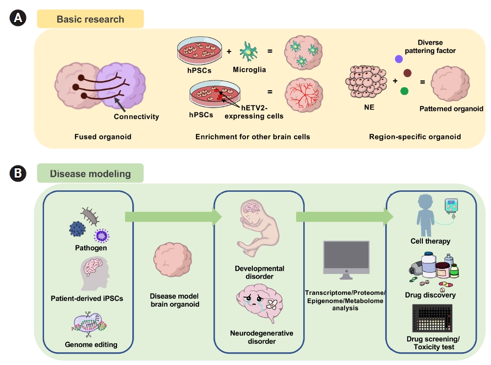
Organoids are among the most accessible and physiologically suitable models to study the differentiation process of stem cells in controlled environments. Brain organoids have been proven to be a high-fidelity platform to reveal the unique identity of stem cells and the niche composition of the surrounding microenvironment [115]. Combined with analyses of genetic information, the transcriptome, and proteins, organoids have significantly contributed to our understanding of brain development, homeostasis maintenance, and key aspects of disease. The process of creating a brain organoid that mirrors human brain development has been utilized in various ways, ranging from basic research tools to applied research. Brain organoids facilitate disease modeling and pathogenesis studies for conditions such as infectious diseases, genetic diseases, and cancer, thereby aiding in the identification of reliable molecular targets [81,116]. When combined with an engineering approach, brain organoids can serve as testing grounds for evaluating drug efficacy and toxicity. The integration of organoid technology with current technologies leads to a variety of subsequent functions and applications, underscoring the versatility of organoids (Fig. 3). These features, coupled with their physiological relevance, position organoids as one of the most exciting and promising technologies recently introduced for studying human brain development, disease, and treatment.

NOTES

Conflict of interest

No potential conflict of interest relevant to this article was reported.

Funding

We thank Hyunjin Lee for the illustration. In-Hyun Park was partly supported by NIH (R01MH118344-01A1). W.Y was supported by the Basic Science Research Program through the National Research Foundation of Korea (NRF) funded by the Ministry of Education(2021R1A6A3A14043824).

Authors’ contributions

Conceptualization: WSY, IHP; Project administration: IHP; Visualization: WSY; Writing-original draft: WSY, FRK; Writing-review & editing: WSY, IHP.

Data availability

Please reach out to the corresponding author to inquire about the availability of data.

Fig. 1.
Development of the neural tube. (A) Three primary vesicles in neural tubes develop into diverse brain areas. Expression of morphogens, fibroblast growth factor (FGF) 8, sonic hedgehog (Shh), and Wnt in neural tubes. (B) Morphogen gradients to specify the developmental axis for dorsal/ventral and rostral/caudal axis.
organoid-2024-4-e1f1.jpg
Fig. 2.
Schematics to generate brain organoids. (A) General steps for generating brain organoids. Methods of culturing organoids in 3-dimensional (3D) and maintaining the organoids in 3D are shown. (B) The unguided approach to produce whole-brain organoids, or cerebral organoids, and the guided approach to produce regionally defined brain organoids. hPSC, human pluripotent stem cell; EB, embryoid body; NE, neural ectoderm.
organoid-2024-4-e1f2.jpg
Fig. 3.
Application and utility of 3-dimensional brain organoids. (A) For basic research, fused organoids or assembloids are used to study neural connectivity. Integrating with non-neuroectodermal cells to investigate microglia and endothelial cells. (B) For disease modeling, brain organoids can be used to study neurodevelopmental and neurodegenerative disorders. hPSC, human pluripotent stem cell; NE, neural ectoderm.
organoid-2024-4-e1f3.jpg
Table 1.
Overview of current telencephalon-related brain organoid protocols
Methodology Type of brain region Type of organoid Extrinsic factors Culture method Extracellular scaffolding Reference
Unguided method Whole brain Cerebral organoid Low bFGF Spinning bioreactor Matrigel [51]
Cerebral organoid Low bFGF Spinning bioreactor Matrigel [52]
Forebrain Cerebral organoid Low bFGF Spinning bioreactor Matrigel [53]
Cerebral organoid Low bFGF Spinning bioreactor Matrigel [54]
Cerebral organoids Low bFGF Spinning bioreactor Matrigel [55]
Guided method Cerebral cortex Cortical spheroid Dorsomorphin, SB-431542, FGF2, EGF Stationary floating - [56,57]
Cortical neuroepithelium IWR1e, SB431542 Stationary floating Matrigel [58]
Cortical organoid Noggin, hDkk1, FGF2 Stationary floating Matrigel on dish [59]
Cortical organoid SB-431542, LDN-193189, XAV939 Orbital shaker - [60]
Forebrain organoid IWR1e, SB431542 Spinning bioreactor Matrigel [61]
Forebrain organoid Wnt3a, Dorsomorphin, A83-01, CHIR99021, SB431542 Miniaturized Spinning Bioreactor Matrigel [62]
Ventral forebrain Ventral organoid IWP-2, SAG Orbital shaker Matrigel [63]
Subpallium spheroids Dorsomorphin, SB431542, IWP-2, SAG Stationary floating - [64]
Medial ganglionic eminence MGE organoid SB431542, LDN193189, XAV939, Shh, Purmorphamine Orbital shaker - [60]
Lateral ganglionic eminence Striatal organoid Dorsomorphin, SB431542, Activin A, IWP-2, SR11237, DAPT Stationary floating - [65]
Hippocampus Hippocampal primordium-like tissue SB431542, IWR1, CHIR99021, and BMP4 Stationary floating - [66]
Hippocampal Spheroids SB-431542, LDN-193189, XAV939, Cyclopamine, CHIR99021 Stationary floating - [67]
Hippocampal organoid Dorsomorphin, A83-01, SB-431542, CHIR-99021, BMP7, Orbital shaker Matrigel [68]
Choroid plexus Choroid plexus-like tissue SB431542, IWR1, CHIR99021, and BMP4 Stationary floating - [66]
Choroid plexus organoid LDN-193189, SB-431542, IWP-2, CHIR-99021, BMP7 Orbital shaker Matrigel [68]
Choroid plexus organoid BMP4, CHIR-99021 Spinning bioreactor Matrigel [69]

bFGF, basic fibroblast growth factor; EGF, epidermal growth factor; IWP-2, inhibitor of Wnt production-2; SAG, smoothened agonist; BMP4/7, bone morphogenetic protein 4/7.

Table 2.
Modeling neurological disorders with telencephalon-related brain organoids
Disease modeling
Type of organoid Disease phenotype of organoid Reference
Disease type Substance/gene
Microcephaly Zika virus Forebrain organoid Decrease of neuronal cell-layer volume, resembling microcephaly [62]
Forebrain organoid Disruption of cortical neurogenesis [83]
Cerebral organoid Perturbed cell fate, a reduction in organoid volume [84]
Cerebral organoid Reduction of proliferative zones, disrupted cortical layers [85]
Cerebral organoid Neural progenitor apoptosis, growth restriction [86]
Cerebral organoid Reduced size and viability, programmed cell death responses. [87]
SARS-CoV-2 virus Cortical organoid - [88]
Cerebral organoid Neuronal cell death, aberrant Tau localization, [89]
Choroid plexus organoid disruption of blood-CSF barrier [90]
HCMV virus Cerebral Organoid Reduction in organoid volume, degeneration of β-tubulin III integrity [91]
Alzheimer's disease APP/PSN1 Hippocampal spheroid Loss of synaptic proteins, increased ratio of intracellular and extracellular Aβ42/Aβ40 peptides [67]
APP Neocortex Aβ aggregation, hyperphosphorylated tau protein, endosome abnormalities [92]
PSN1 Cerebral organoid Higher production of the Aβ protein, increased tau phosphorylation [93]
APOE4 Cerebral organoid Increased levels of Aβ and phosphorylated tau [94]
Rett syndrome MeCP2 Forebrain Organoid Lower expression of neural progenitor, defect of electrophysiological activity [95]
Cortical organoid, MGE organoid Dysregulated gene in neurons and glial cells, abnormal transcription related to synaptic transmission [96]
Autism Spectrum Disorders, FOXG1 Telencephalic organoid Accelerated cell cycle, overproduction of GABAergic inhibitory neurons [59]
CDK5RAP2 Cerebral organoid Premature neuronal differentiation [45]
PTEN Cerebral organoid Delayed neuronal differentiation, expanded VZ and oSVZ, surface expansion and folding [97]
CHD8 Cerebral organoid Dysregulated Wnt/β-catenin signaling, GABAergic interneuron related gene [98]
Miller-Dieker syndrome PAFAH1B1 Cerebral organoid Increase apoptosis/vertical spindle orientation, prolonged mitosis [99]
Timothy syndrome CACNA1C Cortical and subpallium spheroid Abnormal migratory saltation [64]
Fragile X syndrome FMR1 Forebrain organoid Dysregulated neurogenesis, neuronal maturation and neuronal excitability. [100]

SARS-CoV-2, severe acute respiratory syndrome coronavirus 2; HCMV, human cytomegalovirus; APP, amyloid precursor protein; PSN1, presenilin 1; APOE4, apolipoprotein E4; MeCP2, methyl-CpG binding protein 2; FOXG1, forkhead box G1; PTEN, phosphatase and tensin homolog; CDK5RAP2, CDK5 regulatory subunit associated protein 2; CHD8, chromodomain-helicase-DNA-binding protein 8; PAFAH1B1, platelet activating factor acetylhydrolase 1b regulatory subunit 1; CACNA1C, L-type calcium channel Cav1. 2; FMR1, fragile X messenger ribonucleoprotein 1.

References

1. Felten DL, O'Banion MK, Maida MS, Netter FH. Netter's atlas of neuroscience. 3rd ed. Philadelphia: Elsevier; 2016.
2. Haines DE, Mihailoff GA. Fundamental neuroscience for basic and clinical applications. 5th ed. Philadelphia: Elsevier; 2018.
3. Yoshida M, Suda Y, Matsuo I, Miyamoto N, Takeda N, Kuratani S, et al. Emx1 and Emx2 functions in development of dorsal telencephalon. Development 1997;124:101-11.
crossref pmid pdf
4. Sun N, Meng X, Liu Y, Song D, Jiang C, Cai J. Applications of brain organoids in neurodevelopment and neurological diseases. J Biomed Sci 2021;28:30.
crossref pmid pmc pdf
5. Chaves-Coira I, Rodrigo-Angulo ML, Nuñez A. Bilateral pathways from the basal forebrain to sensory cortices may contribute to synchronous sensory processing. Front Neuroanat 2018;12:5.
crossref pmid pmc
6. Jawabri KH, Sharma S. StatPearls. Treasure Island: StatPearls Publishing; 2021 Available from: https://www.ncbi.nlm.nih.gov/books/NBK538496/.
7. Dennis D, Picketts D, Slack RS, Schuurmans C. Forebrain neurogenesis: from embryo to adult. Trends Dev Biol 2016;9:77-90.
pmid pmc
8. El-Baba RM, Schury MP. StatPearls. Treasure Island: StatPearls Publishing; 2021 Available from: https://www.ncbi.nlm.nih.gov/books/NBK554483/.
9. Hoffmann M. The human frontal lobes and frontal network systems: an evolutionary, clinical, and treatment perspective. ISRN Neurol 2013;2013:892459.
crossref pmid pmc pdf
10. Pirau L, Lui F. StatPearls. Treasure Island: StatPearls Publishing; 2021 Available from: https://www.ncbi.nlm.nih.gov/books/NBK532981/.
11. Patel A, Biso G, Fowler JB. StatPearls. Treasure Island: StatPearls Publishing; 2021 Available from: https://www.ncbi.nlm.nih.gov/books/NBK519512/.
12. Williamson PD, Thadani VM, French JA, Darcey TM, Mattson RH, Spencer SS, et al. Medial temporal lobe epilepsy: videotape analysis of objective clinical seizure characteristics. Epilepsia 1998;39:1182-8.
crossref pmid
13. Rehman A, Al Khalili Y. StatPearls. Treasure Island: StatPearls Publishing; 2021 Available from: https://www.ncbi.nlm.nih.gov/books/NBK544320/.
14. Amalnath SD, Kumar S, Deepanjali S, Dutta TK. Balint syndrome. Ann Indian Acad Neurol 2014;17:10-1.
crossref pmid pmc
15. Bisley JW, Goldberg ME. Attention, intention, and priority in the parietal lobe. Annu Rev Neurosci 2010;33:1-21.
crossref pmid pmc
16. Coslett HB, Schwartz MF. The parietal lobe and language. Handb Clin Neurol 2018;151:365-75.
crossref pmid
17. Altabakhi IW, Liang JW. StatPearls. Treasure Island: StatPearls Publishing; 2021 Available from: https://www.ncbi.nlm.nih.gov/books/NBK519528/.
18. Stiles J, Jernigan TL. The basics of brain development. Neuropsychol Rev 2010;20:327-48.
crossref pmid pmc
19. Bond AM, Bhalala OG, Kessler JA. The dynamic role of bone morphogenetic proteins in neural stem cell fate and maturation. Dev Neurobiol 2012;72:1068-84.
crossref pmid pmc
20. Bergquist H. The formation of the front part of the neural tube. Experientia 1964;20:92-3.
crossref pmid pdf
21. Foley AC, Stern CD. Evolution of vertebrate forebrain development: how many different mechanisms? J Anat 2001;199(Pt 1-2):35-52.
crossref pmid pmc
22. Wilson SW, Houart C. Early steps in the development of the forebrain. Dev Cell 2004;6:167-81.
crossref pmid pmc
23. Brafman D, Willert K. Wnt/β-catenin signaling during early vertebrate neural development. Dev Neurobiol 2017;77:1239-59.
crossref pmid pmc pdf
24. Chizhikov VV, Millen KJ. Roof plate-dependent patterning of the vertebrate dorsal central nervous system. Dev Biol 2005;277:287-95.
crossref pmid
25. Montiel JF, Aboitiz F. Pallial patterning and the origin of the isocortex. Front Neurosci 2015;9:377.
crossref pmid pmc
26. Abellán A, Desfilis E, Medina L. Combinatorial expression of Lef1, Lhx2, Lhx5, Lhx9, Lmo3, Lmo4, and Prox1 helps to identify comparable subdivisions in the developing hippocampal formation of mouse and chicken. Front Neuroanat 2014;8:59.
pmid pmc
27. Medina L, Abellán A. Development and evolution of the pallium. Semin Cell Dev Biol 2009;20:698-711.
crossref pmid
28. Medina L, Abellán A, Desfilis E. Evolution of pallial areas and networks involved in sociality: comparison between mammals and sauropsids. Front Physiol 2019;10:894.
crossref pmid pmc
29. Pauly MC, Döbrössy MD, Nikkhah G, Winkler C, Piroth T. Organization of the human fetal subpallium. Front Neuroanat 2014;7:54.
crossref pmid pmc
30. Stern CD. Initial patterning of the central nervous system: how many organizers? Nat Rev Neurosci 2001;2:92-8.
crossref pmid pdf
31. Caronia-Brown G, Yoshida M, Gulden F, Assimacopoulos S, Grove EA. The cortical hem regulates the size and patterning of neocortex. Development 2014;141:2855-65.
crossref pmid pmc pdf
32. Lee SM, Tole S, Grove E, McMahon AP. A local Wnt-3a signal is required for development of the mammalian hippocampus. Development 2000;127:457-67.
crossref pmid pdf
33. Varela-Nallar L, Inestrosa NC. Wnt signaling in the regulation of adult hippocampal neurogenesis. Front Cell Neurosci 2013;7:100.
crossref pmid pmc
34. Galceran J, Miyashita-Lin EM, Devaney E, Rubenstein JL, Grosschedl R. Hippocampus development and generation of dentate gyrus granule cells is regulated by LEF1. Development 2000;127:469-82.
crossref pmid pdf
35. Monuki ES, Porter FD, Walsh CA. Patterning of the dorsal telencephalon and cerebral cortex by a roof plate-Lhx2 pathway. Neuron 2001;32:591-604.
crossref pmid
36. Caronia G, Wilcoxon J, Feldman P, Grove EA. Bone morphogenetic protein signaling in the developing telencephalon controls formation of the hippocampal dentate gyrus and modifies fear-related behavior. J Neurosci 2010;30:6291-301.
crossref pmid pmc
37. Rash BG, Grove EA. Patterning the dorsal telencephalon: a role for sonic hedgehog? J Neurosci 2007;27:11595-603.
crossref pmid pmc
38. Bertrand N, Dahmane N. Sonic hedgehog signaling in forebrain development and its interactions with pathways that modify its effects. Trends Cell Biol 2006;16:597-605.
crossref pmid
39. Peyre E, Silva CG, Nguyen L. Crosstalk between intracellular and extracellular signals regulating interneuron production, migration and integration into the cortex. Front Cell Neurosci 2015;9:129.
crossref pmid pmc
40. Fjodorova M, Noakes Z, Li M. How to make striatal projection neurons. Neurogenesis (Austin) 2015;2:e1100227.
crossref pmid pmc
41. Fuccillo M, Rallu M, McMahon AP, Fishell G. Temporal requirement for hedgehog signaling in ventral telencephalic patterning. Development 2004;131:5031-40.
crossref pmid pdf
42. Lee JY. Normal and disordered formation of the cerebral cortex : normal embryology, related molecules, types of migration, migration disorders. J Korean Neurosurg Soc 2019;62:265-71.
crossref pmid pmc pdf
43. Lehmann R, Lee CM, Shugart EC, Benedetti M, Charo RA, Gartner Z, et al. Human organoids: a new dimension in cell biology. Mol Biol Cell 2019;30:1129-37.
crossref pmid pmc
44. Hofer M, Lutolf MP. Engineering organoids. Nat Rev Mater 2021;6:402-20.
crossref pmid pmc pdf
45. Lancaster MA, Renner M, Martin CA, Wenzel D, Bicknell LS, Hurles ME, et al. Cerebral organoids model human brain development and microcephaly. Nature 2013;501:373-9.
crossref pmid pmc pdf
46. Kim J, Sullivan GJ, Park IH. How well do brain organoids capture your brain? iScience 2021;24:102063.
crossref pmid pmc
47. Tanaka Y, Park IH. Regional specification and complementation with non-neuroectodermal cells in human brain organoids. J Mol Med (Berl) 2021;99:489-500.
crossref pmid pmc pdf
48. Kiral FR, Cakir B, Tanaka Y, Kim J, Yang WS, Wehbe F, et al. Generation of ventralized human thalamic organoids with thalamic reticular nucleus. Cell Stem Cell 2023;30:677-88.
crossref pmid pmc
49. Eiraku M, Watanabe K, Matsuo-Takasaki M, Kawada M, Yonemura S, Matsumura M, et al. Self-organized formation of polarized cortical tissues from ESCs and its active manipulation by extrinsic signals. Cell Stem Cell 2008;3:519-32.
crossref pmid
50. Sasai Y. Next-generation regenerative medicine: organogenesis from stem cells in 3D culture. Cell Stem Cell 2013;12:520-30.
crossref pmid
51. Quadrato G, Nguyen T, Macosko EZ, Sherwood JL, Min Yang S, Berger DR, et al. Cell diversity and network dynamics in photosensitive human brain organoids. Nature 2017;545:48-53.
crossref pmid pmc pdf
52. Hattori N. Cerebral organoids model human brain development and microcephaly. Mov Disord 2014;29:185.
crossref pmid
53. Camp JG, Badsha F, Florio M, Kanton S, Gerber T, Wilsch-Bräuninger M, et al. Human cerebral organoids recapitulate gene expression programs of fetal neocortex development. Proc Natl Acad Sci U S A 2015;112:15672-7.
crossref pmid pmc
54. Mansour AA, Gonçalves JT, Bloyd CW, Li H, Fernandes S, Quang D, et al. An in vivo model of functional and vascularized human brain organoids. Nat Biotechnol 2018;36:432-41.
crossref pmid pmc pdf
55. Renner M, Lancaster MA, Bian S, Choi H, Ku T, Peer A, et al. Self-organized developmental patterning and differentiation in cerebral organoids. EMBO J 2017;36:1316-29.
crossref pmid pmc pdf
56. Paşca AM, Sloan SA, Clarke LE, Tian Y, Makinson CD, Huber N, et al. Functional cortical neurons and astrocytes from human pluripotent stem cells in 3D culture. Nat Methods 2015;12:671-8.
crossref pmid pmc pdf
57. Sloan SA, Darmanis S, Huber N, Khan TA, Birey F, Caneda C, et al. Human astrocyte maturation captured in 3D cerebral cortical spheroids derived from pluripotent stem cells. Neuron 2017;95:779-90.
crossref pmid pmc
58. Kadoshima T, Sakaguchi H, Nakano T, Soen M, Ando S, Eiraku M, et al. Self-organization of axial polarity, inside-out layer pattern, and species-specific progenitor dynamics in human ES cell-derived neocortex. Proc Natl Acad Sci U S A 2013;110:20284-9.
crossref pmid pmc
59. Mariani J, Coppola G, Zhang P, Abyzov A, Provini L, Tomasini L, et al. FOXG1-dependent dysregulation of GABA/glutamate neuron differentiation in autism spectrum disorders. Cell 2015;162:375-90.
crossref pmid pmc
60. Xiang Y, Tanaka Y, Patterson B, Kang YJ, Govindaiah G, Roselaar N, et al. Fusion of regionally specified hPSC-derived organoids models human brain development and interneuron migration. Cell Stem Cell 2017;21:383-98.
crossref pmid pmc
61. Velasco S, Kedaigle AJ, Simmons SK, Nash A, Rocha M, Quadrato G, et al. Individual brain organoids reproducibly form cell diversity of the human cerebral cortex. Nature 2019;570:523-7.
crossref pmid pmc pdf
62. Qian X, Nguyen HN, Song MM, Hadiono C, Ogden SC, Hammack C, et al. Brain-region-specific organoids using mini-bioreactors for modeling ZIKV exposure. Cell 2016;165:1238-54.
pmid pmc
63. Bagley JA, Reumann D, Bian S, Lévi-Strauss J, Knoblich JA. Fused cerebral organoids model interactions between brain regions. Nat Methods 2017;14:743-51.
crossref pmid pmc pdf
64. Birey F, Andersen J, Makinson CD, Islam S, Wei W, Huber N, et al. Assembly of functionally integrated human forebrain spheroids. Nature 2017;545:54-9.
crossref pmid pmc pdf
65. Miura Y, Li MY, Birey F, Ikeda K, Revah O, Thete MV, et al. Generation of human striatal organoids and cortico-striatal assembloids from human pluripotent stem cells. Nat Biotechnol 2020;38:1421-30.
crossref pmid pmc pdf
66. Sakaguchi H, Kadoshima T, Soen M, Narii N, Ishida Y, Ohgushi M, et al. Generation of functional hippocampal neurons from self-organizing human embryonic stem cell-derived dorsomedial telencephalic tissue. Nat Commun 2015;6:8896.
crossref pmid pdf
67. Pomeshchik Y, Klementieva O, Gil J, Martinsson I, Hansen MG, de Vries T, et al. Human iPSC-derived hippocampal spheroids: an innovative tool for stratifying Alzheimer disease patient-specific cellular phenotypes and developing therapies. Stem Cell Reports 2020;15:256-73.
crossref pmid pmc
68. Jacob F, Pather SR, Huang WK, Zhang F, Wong SZ, Zhou H, et al. Human pluripotent stem cell-derived neural cells and brain organoids reveal SARS-CoV-2 neurotropism predominates in choroid plexus epithelium. Cell Stem Cell 2020;27:937-50.
crossref pmid pmc
69. Pellegrini L, Bonfio C, Chadwick J, Begum F, Skehel M, Lancaster MA. Human CNS barrier-forming organoids with cerebrospinal fluid production. Science 2020;369:eaaz5626.
crossref pmid pmc
70. Wattanapanitch M, Klincumhom N, Potirat P, Amornpisutt R, Lorthongpanich C, U-pratya Y, et al. Dual small-molecule targeting of SMAD signaling stimulates human induced pluripotent stem cells toward neural lineages. PLoS One 2014;9:e106952.
crossref pmid pmc
71. Hong YJ, Do JT. Neural lineage differentiation from pluripotent stem cells to mimic human brain tissues. Front Bioeng Biotechnol 2019;7:400.
crossref pmid pmc
72. Jacob F, Schnoll JG, Song H, Ming GL. Building the brain from scratch: engineering region-specific brain organoids from human stem cells to study neural development and disease. Curr Top Dev Biol 2021;142:477-530.
crossref pmid pmc
73. Sidhaye J, Knoblich JA. Brain organoids: an ensemble of bioassays to investigate human neurodevelopment and disease. Cell Death Differ 2021;28:52-67.
crossref pmid pmc pdf
74. Xiang Y, Tanaka Y, Cakir B, Patterson B, Kim KY, Sun P, et al. hESC-derived thalamic organoids form reciprocal projections when fused with cortical organoids. Cell Stem Cell 2019;24:487-97.
crossref pmid pmc
75. Yoon SJ, Elahi LS, Pașca AM, Marton RM, Gordon A, Revah O, et al. Reliability of human cortical organoid generation. Nat Methods 2019;16:75-8.
crossref pmid pmc pdf
76. Hatami M, Conrad S, Naghsh P, Alvarez-Bolado G, Skutella T. Cell-biological requirements for the generation of dentate gyrus granule neurons. Front Cell Neurosci 2018;12:402.
crossref pmid pmc
77. Brady MV, Vaccarino FM. Role of SHH in patterning human pluripotent cells towards ventral forebrain fates. Cells 2021;10:914.
crossref pmid pmc
78. Correction: Sequerra, Goyal et al., “NMDA receptor signaling is important for neural tube formation and for preventing antiepileptic drug-induced neural tube defects”. J Neurosci 2018;38:10399.
crossref pmid pmc
79. Cederquist GY, Asciolla JJ, Tchieu J, Walsh RM, Cornacchia D, Resh MD, et al. Specification of positional identity in forebrain organoids. Nat Biotechnol 2019;37:436-44.
crossref pmid pmc pdf
80. Kretzschmar K, Clevers H. Organoids: modeling development and the stem cell niche in a dish. Dev Cell 2016;38:590-600.
crossref pmid
81. Xu J, Wen Z. Brain organoids: studying human brain development and diseases in a dish. Stem Cells Int 2021;2021:5902824.
crossref pmid pmc pdf
82. Wang H. Modeling neurological diseases with human brain organoids. Front Synaptic Neurosci 2018;10:15.
crossref pmid pmc
83. Yoon KJ, Song G, Qian X, Pan J, Xu D, Rho HS, et al. Zika-virus-encoded NS2A disrupts mammalian cortical neurogenesis by degrading adherens junction proteins. Cell Stem Cell 2017;21:349-58.
crossref pmid pmc
84. Dang J, Tiwari SK, Lichinchi G, Qin Y, Patil VS, Eroshkin AM, et al. Zika virus depletes neural progenitors in human cerebral organoids through activation of the innate immune receptor TLR3. Cell Stem Cell 2016;19:258-65.
crossref pmid pmc
85. Cugola FR, Fernandes IR, Russo FB, Freitas BC, Dias JL, Guimarães KP, et al. The Brazilian Zika virus strain causes birth defects in experimental models. Nature 2016;534:267-71.
crossref pmid pmc pdf
86. Watanabe M, Buth JE, Vishlaghi N, de la Torre-Ubieta L, Taxidis J, Khakh BS, et al. Self-organized cerebral organoids with human-specific features predict effective drugs to combat Zika virus infection. Cell Rep 2017;21:517-32.
crossref pmid pmc
87. Garcez PP, Loiola EC, Madeiro da Costa R, Higa LM, Trindade P, Delvecchio R, et al. Zika virus impairs growth in human neurospheres and brain organoids. Science 2016;352:816-8.
crossref pmid
88. Zhang BZ, Chu H, Han S, Shuai H, Deng J, Hu YF, et al. SARS-CoV-2 infects human neural progenitor cells and brain organoids. Cell Res 2020;30:928-31.
crossref pmid pmc pdf
89. Ramani A, Müller L, Ostermann PN, Gabriel E, Abida-Islam P, Müller-Schiffmann A, et al. SARS-CoV-2 targets neurons of 3D human brain organoids. EMBO J 2020;39:e106230.
pmid pmc
90. Pellegrini L, Albecka A, Mallery DL, Kellner MJ, Paul D, Carter AP, et al. SARS-CoV-2 infects the brain choroid plexus and disrupts the blood-CSF barrier in human brain organoids. Cell Stem Cell 2020;27:951-61.
crossref pmid pmc
91. Brown RM, Rana PS, Jaeger HK, O'Dowd JM, Balemba OB, Fortunato EA. Human cytomegalovirus compromises development of cerebral organoids. J Virol 2019;93:e00957.
crossref pmid pmc pdf
92. Raja WK, Mungenast AE, Lin YT, Ko T, Abdurrob F, Seo J, et al. Self-organizing 3D human neural tissue derived from induced pluripotent stem cells recapitulate Alzheimer's disease phenotypes. PLoS One 2016;11:e0161969.
crossref pmid pmc
93. Gonzalez C, Armijo E, Bravo-Alegria J, Becerra-Calixto A, Mays CE, Soto C. Modeling amyloid beta and tau pathology in human cerebral organoids. Mol Psychiatry 2018;23:2363-74.
crossref pmid pmc pdf
94. Zhao J, Fu Y, Yamazaki Y, Ren Y, Davis MD, Liu CC, et al. APOE4 exacerbates synapse loss and neurodegeneration in Alzheimer's disease patient iPSC-derived cerebral organoids. Nat Commun 2020;11:5540.
crossref pmid pmc pdf
95. Gomes AR, Fernandes TG, Vaz SH, Silva TP, Bekman EP, Xapelli S, et al. Modeling Rett syndrome with human patient-specific forebrain organoids. Front Cell Dev Biol 2020;8:610427.
crossref pmid pmc
96. Xiang Y, Tanaka Y, Patterson B, Hwang SM, Hysolli E, Cakir B, et al. Dysregulation of BRD4 function underlies the functional abnormalities of MeCP2 mutant neurons. Mol Cell 2020;79:84-98.
crossref pmid pmc
97. Li Y, Muffat J, Omer A, Bosch I, Lancaster MA, Sur M, et al. Induction of expansion and folding in human cerebral organoids. Cell Stem Cell 2017;20:385-96.
crossref pmid
98. Wang P, Mokhtari R, Pedrosa E, Kirschenbaum M, Bayrak C, Zheng D, et al. CRISPR/Cas9-mediated heterozygous knockout of the autism gene CHD8 and characterization of its transcriptional networks in cerebral organoids derived from iPS cells. Mol Autism 2017;8:11.
crossref pmid pmc pdf
99. Bershteyn M, Nowakowski TJ, Pollen AA, Di Lullo E, Nene A, Wynshaw-Boris A, et al. Human iPSC-derived cerebral organoids model cellular features of lissencephaly and reveal prolonged mitosis of outer radial glia. Cell Stem Cell 2017;20:435-49.
crossref pmid pmc
100. Kang Y, Zhou Y, Li Y, Han Y, Xu J, Niu W, et al. A human forebrain organoid model of fragile X syndrome exhibits altered neurogenesis and highlights new treatment strategies. Nat Neurosci 2021;24:1377-91.
crossref pmid pmc pdf
101. Kinney JW, Bemiller SM, Murtishaw AS, Leisgang AM, Salazar AM, Lamb BT. Inflammation as a central mechanism in Alzheimer’s disease. Alzheimers Dement (N Y) 2018;4:575-90.
pmid pmc
102. Jacob F, Salinas RD, Zhang DY, Nguyen PT, Schnoll JG, Wong SZ, et al. A patient-derived glioblastoma organoid model and biobank recapitulates inter- and intra-tumoral heterogeneity. Cell 2020;180:188-204.
pmid
103. Jacob F, Ming GL, Song H. Generation and biobanking of patient-derived glioblastoma organoids and their application in CAR T cell testing. Nat Protoc 2020;15:4000-33.
crossref pmid pdf
104. Cho AN, Jin Y, An Y, Kim J, Choi YS, Lee JS, et al. Microfluidic device with brain extracellular matrix promotes structural and functional maturation of human brain organoids. Nat Commun 2021;12:4730.
crossref pmid pmc pdf
105. Silva TP, Sousa-Luís R, Fernandes TG, Bekman EP, Rodrigues CA, Vaz SH, et al. Transcriptome profiling of human pluripotent stem cell-derived cerebellar organoids reveals faster commitment under dynamic conditions. Biotechnol Bioeng 2021;118:2781-803.
crossref pmid pdf
106. Zhu Y, Wang L, Yu H, Yin F, Wang Y, Liu H, et al. In situ generation of human brain organoids on a micropillar array. Lab Chip 2017;17:2941-50.
crossref pmid
107. Reardon S. Lab-grown ‘mini brains’ produce electrical patterns that resemble those of premature babies. Nature 2018;563:453.
crossref pmid pdf
108. Zhao X, Eyo UB, Murugan M, Wu LJ. Microglial interactions with the neurovascular system in physiology and pathology. Dev Neurobiol 2018;78:604-17.
crossref pmid pmc pdf
109. Brown LS, Foster CG, Courtney JM, King NE, Howells DW, Sutherland BA. Pericytes and neurovascular function in the healthy and diseased brain. Front Cell Neurosci 2019;13:282.
crossref pmid pmc
110. Cakir B, Park IH. Getting the right cells. Elife 2022;11:e80373.
crossref pmid pmc pdf
111. Cakir B, Kiral FR, Park IH. Advanced in vitro models: microglia in action. Neuron 2022;110:3444-57.
crossref pmid
112. Cakir B, Xiang Y, Tanaka Y, Kural MH, Parent M, Kang YJ, et al. Engineering of human brain organoids with a functional vascular-like system. Nat Methods 2019;16:1169-75.
crossref pmid pmc
113. Shi Y, Sun L, Wang M, Liu J, Zhong S, Li R, et al. Vascularized human cortical organoids (vOrganoids) model cortical development in vivo. PLoS Biol 2020;18:e3000705.
crossref pmid pmc
114. Ormel PR, Vieira de Sá R, van Bodegraven EJ, Karst H, Harschnitz O, Sneeboer MA, et al. Microglia innately develop within cerebral organoids. Nat Commun 2018;9:4167.
crossref pmid pmc pdf
115. Di Lullo E, Kriegstein AR. The use of brain organoids to investigate neural development and disease. Nat Rev Neurosci 2017;18:573-84.
crossref pmid pmc pdf
116. Trujillo CA, Muotri AR. Brain organoids and the study of neurodevelopment. Trends Mol Med 2018;24:982-90.
crossref pmid pmc


ABOUT
BROWSE ARTICLES
EDITORIAL POLICY
FOR CONTRIBUTORS
Editorial Office
Room 319, Hall 1 of Chonbuk National University Dental College, 20, Geonji-ro, Deokjin-gu, Jeonju 54907, Korea
Tel: +82-63-270-4024    E-mail: editor@j-organoid.org                

Copyright © 2024 by The Organoid Society.

Developed in M2PI

Close layer
prev next